


 Foundational characteristics of cancer include proliferation, angiogenesis, migration, evasion of apoptosis, and cellular immortality. Find key markers for these cellular processes and antibodies to detect them.
 The SUMOplot™ Analysis Program predicts and scores sumoylation sites in your protein. SUMOylation is a post-translational modification involved in various cellular processes, such as nuclear-cytosolic transport, transcriptional regulation, apoptosis, protein stability, response to stress, and progression through the cell cycle.
 The Autophagy Receptor Motif Plotter predicts and scores autophagy receptor binding sites in your protein. Identifying proteins connected to this pathway is critical to understanding the role of autophagy in physiological as well as pathological processes such as development, differentiation, neurodegenerative diseases, stress, infection, and cancer.
Mouse Monoclonal Antibody to AKT3
Purified Mouse Monoclonal Antibody
- SPECIFICATION
- CITATIONS
- PROTOCOLS
- BACKGROUND

Application
| WB, FC, ICC, E |
|---|---|
| Primary Accession | P19022 |
| Reactivity | Human |
| Host | Mouse |
| Clonality | Monoclonal |
| Clone Names | 5H7C3 |
| Isotype | Mouse IgG2a |
| Calculated MW | 55.8kDa |
| Description | This gene encodes a classical cadherin and member of the cadherin superfamily. Alternative splicing results in multiple transcript variants, at least one of which encodes a preproprotein is proteolytically processed to generate a calcium-dependent cell adhesion molecule and glycoprotein. This protein plays a role in the establishment of left-right asymmetry, development of the nervous system and the formation of cartilage and bone.; |
| Immunogen | Purified recombinant fragment of human AKT3 (AA: 37-150) expressed in E. Coli. |
| Formulation | Purified antibody in PBS with 0.05% sodium azide |
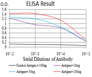
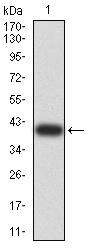
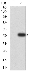
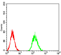
| Application Note | ELISA: 1/10000 WB: 1/500 - 1/2000 ICC: 1/50 - 1/250 FCM: 1/200 - 1/400 |
| Gene ID | 1000 |
|---|---|
| Other Names | CDHN; NCAD; CD325; CDw325 |
| Dilution | WB~~1:1000 FC~~1:10~50 ICC~~N/A E~~N/A |
| Storage | Maintain refrigerated at 2-8°C for up to 6 months. For long term storage store at -20°C in small aliquots to prevent freeze-thaw cycles. |
| Precautions | Mouse Monoclonal Antibody to AKT3 is for research use only and not for use in diagnostic or therapeutic procedures. |
| Name | CDH2 |
|---|---|
| Synonyms | CDHN, NCAD |
| Function | Calcium-dependent cell adhesion protein; preferentially mediates homotypic cell-cell adhesion by dimerization with a CDH2 chain from another cell. Cadherins may thus contribute to the sorting of heterogeneous cell types. Acts as a regulator of neural stem cells quiescence by mediating anchorage of neural stem cells to ependymocytes in the adult subependymal zone: upon cleavage by MMP24, CDH2-mediated anchorage is affected, leading to modulate neural stem cell quiescence. Plays a role in cell-to-cell junction formation between pancreatic beta cells and neural crest stem (NCS) cells, promoting the formation of processes by NCS cells (By similarity). Required for proper neurite branching. Required for pre- and postsynaptic organization (By similarity). CDH2 may be involved in neuronal recognition mechanism. In hippocampal neurons, may regulate dendritic spine density. |
| Cellular Location | Cell membrane; Single-pass type I membrane protein. Cell membrane, sarcolemma {ECO:0000250|UniProtKB:P15116}. Cell junction. Cell surface {ECO:0000250|UniProtKB:P15116}. Cell junction, desmosome {ECO:0000250|UniProtKB:P15116}. Cell junction, adherens junction {ECO:0000250|UniProtKB:P15116}. Note=Colocalizes with TMEM65 at the intercalated disk in cardiomyocytes. Colocalizes with OBSCN at the intercalated disk and at sarcolemma in cardiomyocytes {ECO:0000250|UniProtKB:P15116} |

Thousands of laboratories across the world have published research that depended on the performance of antibodies from Abcepta to advance their research. Check out links to articles that cite our products in major peer-reviewed journals, organized by research category.
info@abcepta.com, and receive a free "I Love Antibodies" mug.

Provided below are standard protocols that you may find useful for product applications.
References
1.Mol Med Rep. 2015 Aug;12(2):2999-3006. ; 2.BMC Cancer. 2013 Jun 26;13:309. ;
If you have used an Abcepta product and would like to share how it has performed, please click on the "Submit Review" button and provide the requested information. Our staff will examine and post your review and contact you if needed.
If you have any additional inquiries please email technical services at tech@abcepta.com.
Ordering Information
Other Products
Shipping Information
