


 Foundational characteristics of cancer include proliferation, angiogenesis, migration, evasion of apoptosis, and cellular immortality. Find key markers for these cellular processes and antibodies to detect them.
 The SUMOplot™ Analysis Program predicts and scores sumoylation sites in your protein. SUMOylation is a post-translational modification involved in various cellular processes, such as nuclear-cytosolic transport, transcriptional regulation, apoptosis, protein stability, response to stress, and progression through the cell cycle.
 The Autophagy Receptor Motif Plotter predicts and scores autophagy receptor binding sites in your protein. Identifying proteins connected to this pathway is critical to understanding the role of autophagy in physiological as well as pathological processes such as development, differentiation, neurodegenerative diseases, stress, infection, and cancer.
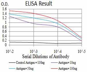
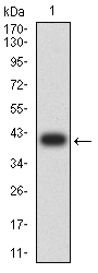
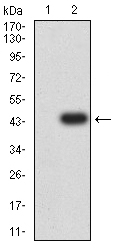
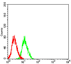
GRIN3B
Purified Mouse Monoclonal Antibody
- SPECIFICATION
- CITATIONS
- PROTOCOLS
- BACKGROUND

Application
| WB, IHC, ICC, E |
|---|---|
| Primary Accession | O60391 |
| Reactivity | Human |
| Host | Mouse |
| Clonality | Monoclonal |
| Clone Names | 2A8E11 |
| Isotype | Mouse IgG2a |
| Calculated MW | 113kDa |
| Immunogen | Purified recombinant fragment of human GRIN3B (AA: 135-276) expressed in E. Coli. |
| Formulation | Purified antibody in PBS with 0.05% sodium azide |
| Gene ID | 116444 |
|---|---|
| Other Names | NR3B; GluN3B |
| Dilution | WB~~ 1/500 - 1/2000 IHC~~1:100~500 ICC~~N/A E~~ 1/10000 |
| Storage | Maintain refrigerated at 2-8°C for up to 6 months. For long term storage store at -20°C in small aliquots to prevent freeze-thaw cycles. |
| Precautions | GRIN3B is for research use only and not for use in diagnostic or therapeutic procedures. |
| Name | GRIN3B (HGNC:16768) |
|---|---|
| Function | Component of a non-conventional N-methyl-D-aspartate (NMDA) receptors (NMDARs) that function as heterotetrameric, ligand-gated cation channels with low calcium permeability and low voltage-dependent block by Mg(2+) (By similarity). Forms glutamatergic receptor complexes with GluN1 and GluN2 subunits which are activated by glycine binding to the GluN1 and GluN3 subunits and L-glutamate binding to GluN2 subunits (By similarity). Forms excitatory glycinergic receptor complexes with GluN1 alone which are activated by glycine binding to the GluN1 and GluN3 subunits. GluN3B subunit also binds D-serine and, in the absence of glycine, activates glycinergic receptor complexes, but with lower efficacy than glycine (By similarity). Each GluN3 subunit confers differential attributes to channel properties, including activation, deactivation and desensitization kinetics, pH sensitivity, Ca2(+) permeability, and binding to allosteric modulators (By similarity). |
| Cellular Location | Cell membrane {ECO:0000250|UniProtKB:Q91ZU9}; Multi-pass membrane protein {ECO:0000250|UniProtKB:Q13224} Postsynaptic cell membrane {ECO:0000250|UniProtKB:Q91ZU9} Note=Requires the presence of GRIN1 to be targeted at the plasma membrane. {ECO:0000250|UniProtKB:Q91ZU9} |

Thousands of laboratories across the world have published research that depended on the performance of antibodies from Abcepta to advance their research. Check out links to articles that cite our products in major peer-reviewed journals, organized by research category.
info@abcepta.com, and receive a free "I Love Antibodies" mug.

Provided below are standard protocols that you may find useful for product applications.
References
1.PLoS One. 2015 Mar 13;10(3):e0116319.2.Psychiatry Res. 2014 Aug 30;218(3):356-8.
If you have used an Abcepta product and would like to share how it has performed, please click on the "Submit Review" button and provide the requested information. Our staff will examine and post your review and contact you if needed.
If you have any additional inquiries please email technical services at tech@abcepta.com.

Ordering Information
Shipping Information
