


 Foundational characteristics of cancer include proliferation, angiogenesis, migration, evasion of apoptosis, and cellular immortality. Find key markers for these cellular processes and antibodies to detect them.
 The SUMOplot™ Analysis Program predicts and scores sumoylation sites in your protein. SUMOylation is a post-translational modification involved in various cellular processes, such as nuclear-cytosolic transport, transcriptional regulation, apoptosis, protein stability, response to stress, and progression through the cell cycle.
 The Autophagy Receptor Motif Plotter predicts and scores autophagy receptor binding sites in your protein. Identifying proteins connected to this pathway is critical to understanding the role of autophagy in physiological as well as pathological processes such as development, differentiation, neurodegenerative diseases, stress, infection, and cancer.
ALDH1A1
Purified Mouse Monoclonal Antibody
- SPECIFICATION
- CITATIONS
- PROTOCOLS
- BACKGROUND

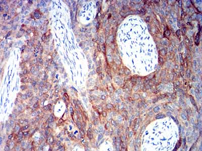
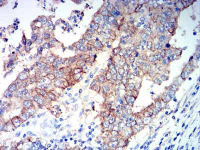
Application
| WB, IHC, ICC, E |
|---|---|
| Primary Accession | P00352 |
| Reactivity | Human |
| Host | Mouse |
| Clonality | Monoclonal |
| Clone Names | 2G1G4 |
| Isotype | Mouse IgG1 |
| Calculated MW | 54.9kDa |
| Immunogen | Purified recombinant fragment of human ALDH1A1 (AA: 1-110) expressed in E. Coli. |
| Formulation | Purified antibody in PBS with 0.05% sodium azide |
| Gene ID | 216 |
|---|---|
| Other Names | ALDC; ALDH1; HEL-9; HEL12; PUMB1; ALDH11; RALDH1; ALDH-E1; HEL-S-53e |
| Dilution | WB~~ 1/500 - 1/2000 IHC~~ 1/200 - 1/1000 ICC~~N/A E~~ 1/10000 |
| Storage | Maintain refrigerated at 2-8°C for up to 6 months. For long term storage store at -20°C in small aliquots to prevent freeze-thaw cycles. |
| Precautions | ALDH1A1 is for research use only and not for use in diagnostic or therapeutic procedures. |
| Name | ALDH1A1 (HGNC:402) |
|---|---|
| Function | Cytosolic dehydrogenase that catalyzes the irreversible oxidation of a wide range of aldehydes to their corresponding carboxylic acid (PubMed:12941160, PubMed:15623782, PubMed:17175089, PubMed:19296407, PubMed:25450233, PubMed:26373694). Functions downstream of retinol dehydrogenases and catalyzes the oxidation of retinaldehyde into retinoic acid, the second step in the oxidation of retinol/vitamin A into retinoic acid (By similarity). This pathway is crucial to control the levels of retinol and retinoic acid, two important molecules which excess can be teratogenic and cytotoxic (By similarity). Also oxidizes aldehydes resulting from lipid peroxidation like (E)-4-hydroxynon-2-enal/HNE, malonaldehyde and hexanal that form protein adducts and are highly cytotoxic. By participating for instance to the clearance of (E)-4-hydroxynon-2-enal/HNE in the lens epithelium prevents the formation of HNE-protein adducts and lens opacification (PubMed:12941160, PubMed:15623782, PubMed:19296407). Also functions downstream of fructosamine-3-kinase in the fructosamine degradation pathway by catalyzing the oxidation of 3-deoxyglucosone, the carbohydrate product of fructosamine 3-phosphate decomposition, which is itself a potent glycating agent that may react with lysine and arginine side-chains of proteins (PubMed:17175089). Also has an aminobutyraldehyde dehydrogenase activity and is probably part of an alternative pathway for the biosynthesis of GABA/4-aminobutanoate in midbrain, thereby playing a role in GABAergic synaptic transmission (By similarity). |
| Cellular Location | Cytoplasm, cytosol. Cell projection, axon {ECO:0000250|UniProtKB:P24549} |
| Tissue Location | Expressed by erythrocytes (at protein level). |

Thousands of laboratories across the world have published research that depended on the performance of antibodies from Abcepta to advance their research. Check out links to articles that cite our products in major peer-reviewed journals, organized by research category.
info@abcepta.com, and receive a free "I Love Antibodies" mug.

Provided below are standard protocols that you may find useful for product applications.
References
1.Oncotarget. 2015 Dec 1;6(38):41360-9.2.Biomark Med. 2015;9(8):777-90.
If you have used an Abcepta product and would like to share how it has performed, please click on the "Submit Review" button and provide the requested information. Our staff will examine and post your review and contact you if needed.
If you have any additional inquiries please email technical services at tech@abcepta.com.

Ordering Information
Other Products
Shipping Information
