


 Foundational characteristics of cancer include proliferation, angiogenesis, migration, evasion of apoptosis, and cellular immortality. Find key markers for these cellular processes and antibodies to detect them.
 The SUMOplot™ Analysis Program predicts and scores sumoylation sites in your protein. SUMOylation is a post-translational modification involved in various cellular processes, such as nuclear-cytosolic transport, transcriptional regulation, apoptosis, protein stability, response to stress, and progression through the cell cycle.
 The Autophagy Receptor Motif Plotter predicts and scores autophagy receptor binding sites in your protein. Identifying proteins connected to this pathway is critical to understanding the role of autophagy in physiological as well as pathological processes such as development, differentiation, neurodegenerative diseases, stress, infection, and cancer.
MLXIP Antibody (N-term)
Affinity Purified Rabbit Polyclonal Antibody (Pab)
- SPECIFICATION
- CITATIONS
- PROTOCOLS
- BACKGROUND

Application
| IHC-P, WB, E |
|---|---|
| Primary Accession | Q9HAP2 |
| Other Accession | Q2VPU4, NP_055753.3 |
| Reactivity | Human, Mouse |
| Host | Rabbit |
| Clonality | Polyclonal |
| Isotype | Rabbit IgG |
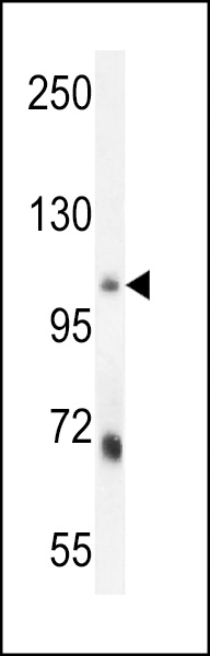
| Calculated MW | 101185 Da |
| Antigen Region | 72-100 aa |
| Gene ID | 22877 |
|---|---|
| Other Names | MLX-interacting protein, Class E basic helix-loop-helix protein 36, bHLHe36, Transcriptional activator MondoA, MLXIP (HGNC:17055) |
| Target/Specificity | This MLXIP antibody is generated from rabbits immunized with a KLH conjugated synthetic peptide between 72-100 amino acids from the N-terminal region of human MLXIP. |
| Dilution | IHC-P~~1:50~100 WB~~1:1000 E~~Use at an assay dependent concentration. |
| Format | Purified polyclonal antibody supplied in PBS with 0.09% (W/V) sodium azide. This antibody is purified through a protein A column, followed by peptide affinity purification. |
| Storage | Maintain refrigerated at 2-8°C for up to 2 weeks. For long term storage store at -20°C in small aliquots to prevent freeze-thaw cycles. |
| Precautions | MLXIP Antibody (N-term) is for research use only and not for use in diagnostic or therapeutic procedures. |
| Name | MLXIP (HGNC:17055) |
|---|---|
| Function | Binds DNA as a heterodimer with MLX and activates transcription. Binds to the canonical E box sequence 5'-CACGTG-3'. Plays a role in transcriptional activation of glycolytic target genes. Involved in glucose-responsive gene regulation. |
| Cellular Location | Cytoplasm. Nucleus {ECO:0000255|PROSITE- ProRule:PRU00981, ECO:0000269|PubMed:12446771, ECO:0000269|PubMed:16782875}. Mitochondrion outer membrane Note=Predominantly cytoplasmic but shuttles between cytoplasm and nucleus when associated with MLX. Also associates with the outer mitochondrial membrane and may shuttle between the outer mitochondrial membrane and the nucleus. {ECO:0000250|UniProtKB:Q2VPU4, ECO:0000269|PubMed:12446771, ECO:0000269|PubMed:16782875} |
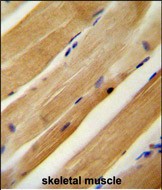
| Tissue Location | Widely expressed in adult tissues. Most abundant in skeletal muscle. |

Thousands of laboratories across the world have published research that depended on the performance of antibodies from Abcepta to advance their research. Check out links to articles that cite our products in major peer-reviewed journals, organized by research category.
info@abcepta.com, and receive a free "I Love Antibodies" mug.

Provided below are standard protocols that you may find useful for product applications.
Background
MONDOA forms heterodimers with MLX (MIM 602976) that can bind to and activate transcription from CACGTG E boxes (Billin et al., 2000 [PubMed 11073985]).
References
Peterson, C.W., et al. Mol. Cell. Biol. 30(12):2887-2895(2010)
Kaadige, M.R., et al. Proc. Natl. Acad. Sci. U.S.A. 106(35):14878-14883(2009)
Stoltzman, C.A., et al. Proc. Natl. Acad. Sci. U.S.A. 105(19):6912-6917(2008)
Sans, C.L., et al. Mol. Cell. Biol. 26(13):4863-4871(2006)
Bornhauser, B.C., et al. J. Biol. Chem. 278(37):35412-35420(2003)
If you have used an Abcepta product and would like to share how it has performed, please click on the "Submit Review" button and provide the requested information. Our staff will examine and post your review and contact you if needed.
If you have any additional inquiries please email technical services at tech@abcepta.com.
Ordering Information
Other Products
Shipping Information
