


 Foundational characteristics of cancer include proliferation, angiogenesis, migration, evasion of apoptosis, and cellular immortality. Find key markers for these cellular processes and antibodies to detect them.
 The SUMOplot™ Analysis Program predicts and scores sumoylation sites in your protein. SUMOylation is a post-translational modification involved in various cellular processes, such as nuclear-cytosolic transport, transcriptional regulation, apoptosis, protein stability, response to stress, and progression through the cell cycle.
 The Autophagy Receptor Motif Plotter predicts and scores autophagy receptor binding sites in your protein. Identifying proteins connected to this pathway is critical to understanding the role of autophagy in physiological as well as pathological processes such as development, differentiation, neurodegenerative diseases, stress, infection, and cancer.
COBL Antibody (N-term)
Affinity Purified Rabbit Polyclonal Antibody (Pab)
- SPECIFICATION
- CITATIONS
- PROTOCOLS
- BACKGROUND

Application
| WB, E |
|---|---|
| Primary Accession | O75128 |
| Other Accession | D3ZUI5, NP_056013.2 |
| Reactivity | Human, Mouse |
| Predicted | Rat |
| Host | Rabbit |
| Clonality | Polyclonal |
| Isotype | Rabbit IgG |
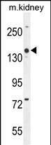
| Calculated MW | 135617 Da |
| Antigen Region | 184-211 aa |
| Gene ID | 23242 |
|---|---|
| Other Names | Protein cordon-bleu, COBL, KIAA0633 |
| Target/Specificity | This COBL antibody is generated from rabbits immunized with a KLH conjugated synthetic peptide between 184-211 amino acids from the N-terminal region of human COBL. |
| Dilution | WB~~1:1000 E~~Use at an assay dependent concentration. |
| Format | Purified polyclonal antibody supplied in PBS with 0.09% (W/V) sodium azide. This antibody is purified through a protein A column, followed by peptide affinity purification. |
| Storage | Maintain refrigerated at 2-8°C for up to 2 weeks. For long term storage store at -20°C in small aliquots to prevent freeze-thaw cycles. |
| Precautions | COBL Antibody (N-term) is for research use only and not for use in diagnostic or therapeutic procedures. |
| Name | COBL |
|---|---|
| Synonyms | KIAA0633 |
| Function | Plays an important role in the reorganization of the actin cytoskeleton. Regulates neuron morphogenesis and increases branching of axons and dendrites. Regulates dendrite branching in Purkinje cells (By similarity). Binds to and sequesters actin monomers (G actin). Nucleates actin polymerization by assembling three actin monomers in cross-filament orientation and thereby promotes growth of actin filaments at the barbed end. Can also mediate actin depolymerization at barbed ends and severing of actin filaments. Promotes formation of cell ruffles. |
| Cellular Location | Cell membrane; Peripheral membrane protein; Cytoplasmic side. Cytoplasm, cytoskeleton. Cell projection, ruffle Cytoplasm. Note=Recruited to the cell membrane via interaction with PACSIN1. Colocalizes with the actin cytoskeleton Detected throughout the neuron cell body, as well as in axons and dendrites (By similarity). |

Thousands of laboratories across the world have published research that depended on the performance of antibodies from Abcepta to advance their research. Check out links to articles that cite our products in major peer-reviewed journals, organized by research category.
info@abcepta.com, and receive a free "I Love Antibodies" mug.

Provided below are standard protocols that you may find useful for product applications.
References
Rose, J.E., et al. Mol. Med. 16 (7-8), 247-253 (2010) :
Trynka, G., et al. Gut 58(8):1078-1083(2009)
Barrett, J.C., et al. Nat. Genet. (2009) In press :
de Krom, M., et al. Biol. Psychiatry 65(7):625-630(2009)
Ballif, B.A., et al. Mol. Cell Proteomics 3(11):1093-1101(2004)
If you have used an Abcepta product and would like to share how it has performed, please click on the "Submit Review" button and provide the requested information. Our staff will examine and post your review and contact you if needed.
If you have any additional inquiries please email technical services at tech@abcepta.com.
Ordering Information
Other Products
Shipping Information
