


 Foundational characteristics of cancer include proliferation, angiogenesis, migration, evasion of apoptosis, and cellular immortality. Find key markers for these cellular processes and antibodies to detect them.
 The SUMOplot™ Analysis Program predicts and scores sumoylation sites in your protein. SUMOylation is a post-translational modification involved in various cellular processes, such as nuclear-cytosolic transport, transcriptional regulation, apoptosis, protein stability, response to stress, and progression through the cell cycle.
 The Autophagy Receptor Motif Plotter predicts and scores autophagy receptor binding sites in your protein. Identifying proteins connected to this pathway is critical to understanding the role of autophagy in physiological as well as pathological processes such as development, differentiation, neurodegenerative diseases, stress, infection, and cancer.
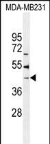
B4GALT6 Antibody (C-term)
Affinity Purified Rabbit Polyclonal Antibody (Pab)
- SPECIFICATION
- CITATIONS
- PROTOCOLS
- BACKGROUND

Application
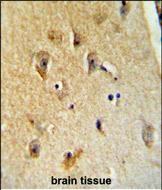
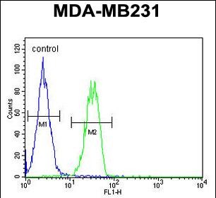
| FC, IHC-P, WB, E |
|---|---|
| Primary Accession | Q9UBX8 |
| Other Accession | Q9WVK5, NP_004766.2 |
| Reactivity | Human |
| Predicted | Mouse |
| Host | Rabbit |
| Clonality | Polyclonal |
| Isotype | Rabbit IgG |
| Calculated MW | 44914 Da |
| Antigen Region | 319-346 aa |
| Gene ID | 9331 |
|---|---|
| Other Names | Beta-1, 4-galactosyltransferase 6, Beta-1, 4-GalTase 6, Beta4Gal-T6, b4Gal-T6, 241-, UDP-Gal:beta-GlcNAc beta-1, 4-galactosyltransferase 6, UDP-galactose:beta-N-acetylglucosamine beta-1, 4-galactosyltransferase 6, Glucosylceramide beta-1, 4-galactosyltransferase, Lactosylceramide synthase, LacCer synthase, UDP-Gal:glucosylceramide beta-1, 4-galactosyltransferase, B4GALT6 |
| Target/Specificity | This B4GALT6 antibody is generated from rabbits immunized with a KLH conjugated synthetic peptide between 319-346 amino acids from the C-terminal region of human B4GALT6. |
| Dilution | FC~~1:10~50 IHC-P~~1:50~100 WB~~1:1000 E~~Use at an assay dependent concentration. |
| Format | Purified polyclonal antibody supplied in PBS with 0.09% (W/V) sodium azide. This antibody is purified through a protein A column, followed by peptide affinity purification. |
| Storage | Maintain refrigerated at 2-8°C for up to 2 weeks. For long term storage store at -20°C in small aliquots to prevent freeze-thaw cycles. |
| Precautions | B4GALT6 Antibody (C-term) is for research use only and not for use in diagnostic or therapeutic procedures. |
| Name | B4GALT6 (HGNC:929) |
|---|---|
| Function | Catalyzes the synthesis of lactosylceramide (LacCer) via the transfer of galactose from UDP-galactose to glucosylceramide (GlcCer) (PubMed:1551920, PubMed:24498430, PubMed:3099851). LacCer is the starting point in the biosynthesis of all gangliosides (membrane-bound glycosphingolipids) which play pivotal roles in the CNS including neuronal maturation and axonal and myelin formation (By similarity). |
| Cellular Location | Golgi apparatus, Golgi stack membrane {ECO:0000250|UniProtKB:P15291}; Single-pass type II membrane protein Note=Trans cisternae of Golgi stack. {ECO:0000250|UniProtKB:P15291} |
| Tissue Location | High expression in brain and adrenal gland, lower in liver, lung, colon and peripheral white blood cells |

Thousands of laboratories across the world have published research that depended on the performance of antibodies from Abcepta to advance their research. Check out links to articles that cite our products in major peer-reviewed journals, organized by research category.
info@abcepta.com, and receive a free "I Love Antibodies" mug.

Provided below are standard protocols that you may find useful for product applications.
Background
This gene is one of seven beta-1,4-galactosyltransferase (beta4GalT) genes. They encode type II membrane-bound glycoproteins that appear to have exclusive specificity for the donor substrate UDP-galactose; all transfer galactose in a beta1,4 linkage to similar acceptor sugars: GlcNAc, Glc, and Xyl. Each beta4GalT has a distinct function in the biosynthesis of different glycoconjugates and saccharide structures. As type II membrane proteins, they have an N-terminal hydrophobic signal sequence that directs the protein to the Golgi apparatus and which then remains uncleaved to function as a transmembrane anchor. By sequence similarity, the beta4GalTs form four groups: beta4GalT1 and beta4GalT2, beta4GalT3 and beta4GalT4, beta4GalT5 and beta4GalT6, and beta4GalT7. The enzyme encoded by this gene is a lactosylceramide synthase important for glycolipid biosynthesis.
References
Landers, J.E., et al. Proc. Natl. Acad. Sci. U.S.A. 106(22):9004-9009(2009) Gevaert, K., et al. Nat. Biotechnol. 21(5):566-569(2003) Fan, Y., et al. DNA Seq. 13(1):1-8(2002) Amado, M., et al. Biochim. Biophys. Acta 1473(1):35-53(1999) Takizawa, M., et al. Biochim. Biophys. Acta 1438(2):301-304(1999)
If you have used an Abcepta product and would like to share how it has performed, please click on the "Submit Review" button and provide the requested information. Our staff will examine and post your review and contact you if needed.
If you have any additional inquiries please email technical services at tech@abcepta.com.
Ordering Information
Shipping Information
