


 Foundational characteristics of cancer include proliferation, angiogenesis, migration, evasion of apoptosis, and cellular immortality. Find key markers for these cellular processes and antibodies to detect them.
 The SUMOplot™ Analysis Program predicts and scores sumoylation sites in your protein. SUMOylation is a post-translational modification involved in various cellular processes, such as nuclear-cytosolic transport, transcriptional regulation, apoptosis, protein stability, response to stress, and progression through the cell cycle.
 The Autophagy Receptor Motif Plotter predicts and scores autophagy receptor binding sites in your protein. Identifying proteins connected to this pathway is critical to understanding the role of autophagy in physiological as well as pathological processes such as development, differentiation, neurodegenerative diseases, stress, infection, and cancer.
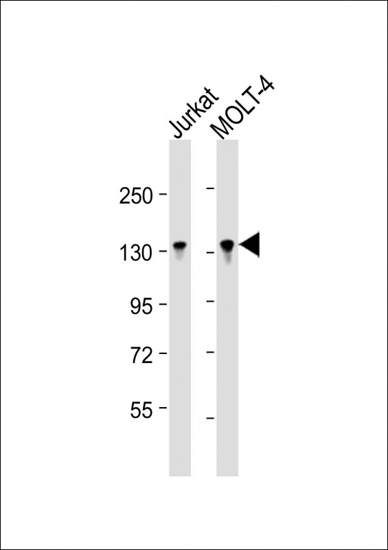
BCL11B Antibody (Center)
Affinity Purified Rabbit Polyclonal Antibody (Pab)
- SPECIFICATION
- CITATIONS
- PROTOCOLS
- BACKGROUND

Application
| WB, E |
|---|---|
| Primary Accession | Q9C0K0 |
| Other Accession | Q99PV8, NP_612808.1, NP_075049.1 |
| Reactivity | Human |
| Predicted | Mouse |
| Host | Rabbit |
| Clonality | Polyclonal |
| Isotype | Rabbit IgG |
| Calculated MW | 95519 Da |
| Antigen Region | 307-334 aa |
| Gene ID | 64919 |
|---|---|
| Other Names | B-cell lymphoma/leukemia 11B, BCL-11B, B-cell CLL/lymphoma 11B, COUP-TF-interacting protein 2, Radiation-induced tumor suppressor gene 1 protein, hRit1, BCL11B, CTIP2, RIT1 |
| Target/Specificity | This BCL11B antibody is generated from rabbits immunized with a KLH conjugated synthetic peptide between 307-334 amino acids from the Central region of human BCL11B. |
| Dilution | WB~~1:16000 E~~Use at an assay dependent concentration. |
| Format | Purified polyclonal antibody supplied in PBS with 0.09% (W/V) sodium azide. This antibody is purified through a protein A column, followed by peptide affinity purification. |
| Storage | Maintain refrigerated at 2-8°C for up to 2 weeks. For long term storage store at -20°C in small aliquots to prevent freeze-thaw cycles. |
| Precautions | BCL11B Antibody (Center) is for research use only and not for use in diagnostic or therapeutic procedures. |
| Name | BCL11B |
|---|---|
| Synonyms | CTIP2, RIT1 |
| Function | Key regulator of both differentiation and survival of T- lymphocytes during thymocyte development in mammals. Essential in controlling the responsiveness of hematopoietic stem cells to chemotactic signals by modulating the expression of the receptors CCR7 and CCR9, which direct the movement of progenitor cells from the bone marrow to the thymus (PubMed:27959755). Is a regulator of IL2 promoter and enhances IL2 expression in activated CD4(+) T-lymphocytes (PubMed:16809611). Tumor-suppressor that represses transcription through direct, TFCOUP2-independent binding to a GC-rich response element (By similarity). May also function in the P53-signaling pathway (By similarity). |
| Cellular Location | Nucleus. |
| Tissue Location | Highly expressed in brain and in malignant T-cell lines derived from patients with adult T-cell leukemia/lymphoma |

Thousands of laboratories across the world have published research that depended on the performance of antibodies from Abcepta to advance their research. Check out links to articles that cite our products in major peer-reviewed journals, organized by research category.
info@abcepta.com, and receive a free "I Love Antibodies" mug.

Provided below are standard protocols that you may find useful for product applications.
Background
This gene encodes a C2H2-type zinc finger protein and is closely related to BCL11A, a gene whose translocation may be associated with B-cell malignancies. The specific function of this gene has not yet been determined. Two alternatively spliced transcript variants, which encode distinct isoforms, have been reported.
References
Ganguli-Indra, G., et al. Exp. Dermatol. 18(11):994-996(2009)
Cherrier, T., et al. Oncogene 28(38):3380-3389(2009)
Ganguli-Indra, G., et al. PLoS ONE 4 (4), E5367 (2009) :
Cismasiu, V.B., et al. Virology 380(2):173-181(2008)
Desplats, P.A., et al. Neurobiol. Dis. 31(3):298-308(2008)
If you have used an Abcepta product and would like to share how it has performed, please click on the "Submit Review" button and provide the requested information. Our staff will examine and post your review and contact you if needed.
If you have any additional inquiries please email technical services at tech@abcepta.com.
Ordering Information
Other Products
Shipping Information
