


 Foundational characteristics of cancer include proliferation, angiogenesis, migration, evasion of apoptosis, and cellular immortality. Find key markers for these cellular processes and antibodies to detect them.
 The SUMOplot™ Analysis Program predicts and scores sumoylation sites in your protein. SUMOylation is a post-translational modification involved in various cellular processes, such as nuclear-cytosolic transport, transcriptional regulation, apoptosis, protein stability, response to stress, and progression through the cell cycle.
 The Autophagy Receptor Motif Plotter predicts and scores autophagy receptor binding sites in your protein. Identifying proteins connected to this pathway is critical to understanding the role of autophagy in physiological as well as pathological processes such as development, differentiation, neurodegenerative diseases, stress, infection, and cancer.
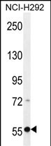
RAD26 Antibody (Center)
Affinity Purified Rabbit Polyclonal Antibody (Pab)
- SPECIFICATION
- CITATIONS
- PROTOCOLS
- BACKGROUND

Application
| WB, E |
|---|---|
| Primary Accession | Q5T890 |
| Other Accession | NP_001010895.1 |
| Reactivity | Human |
| Host | Rabbit |
| Clonality | Polyclonal |
| Isotype | Rabbit IgG |
| Calculated MW | 176051 Da |
| Antigen Region | 226-254 aa |
| Gene ID | 375748 |
|---|---|
| Other Names | DNA excision repair protein ERCC-6-like 2, 364-, DNA repair and recombination protein RAD26-like, ERCC6L2, C9orf102, RAD26L |
| Target/Specificity | This RAD26 antibody is generated from rabbits immunized with a KLH conjugated synthetic peptide between 226-254 amino acids from the Central region of human RAD26. |
| Dilution | WB~~1:1000 E~~Use at an assay dependent concentration. |
| Format | Purified polyclonal antibody supplied in PBS with 0.09% (W/V) sodium azide. This antibody is purified through a protein A column, followed by peptide affinity purification. |
| Storage | Maintain refrigerated at 2-8°C for up to 2 weeks. For long term storage store at -20°C in small aliquots to prevent freeze-thaw cycles. |
| Precautions | RAD26 Antibody (Center) is for research use only and not for use in diagnostic or therapeutic procedures. |
| Name | ERCC6L2 (HGNC:26922) |
|---|---|
| Synonyms | C9orf102, RAD26L |
| Function | Promotes double-strand break (DSB) end-joining and facilitates programmed recombination by controlling how DNA ends are joined in a spatially oriented manner during repair (By similarity). Also plays a role in DNA repair by restricting DNA end resection in double strand break (DSB) repair (PubMed:24507776, PubMed:37014751). Facilitates replication of complex DNA regions and regulates the maintenance of chromatin structure (PubMed:37014751). |
| Cellular Location | Nucleus. Cytoplasm, cytoskeleton, microtubule organizing center, centrosome Mitochondrion. Chromosome, centromere. Note=Colocalizes with NEK6 in the centrosome (PubMed:20873783). In response to DNA damage, translocates from the cytosol to mitochondria and nucleus in a reactive oxygen species (ROS)-dependent manner (PubMed:24507776). Centromeric localization is facilitated by its interaction with PCNA (PubMed:37014751). |
| Tissue Location | Expressed in bone marrow (at protein level). |

Thousands of laboratories across the world have published research that depended on the performance of antibodies from Abcepta to advance their research. Check out links to articles that cite our products in major peer-reviewed journals, organized by research category.
info@abcepta.com, and receive a free "I Love Antibodies" mug.

Provided below are standard protocols that you may find useful for product applications.
References
Lamesch, P., et al. Genomics 89(3):307-315(2007)
Venter, J.C., et al. Science 291(5507):1304-1351(2001)
If you have used an Abcepta product and would like to share how it has performed, please click on the "Submit Review" button and provide the requested information. Our staff will examine and post your review and contact you if needed.
If you have any additional inquiries please email technical services at tech@abcepta.com.
Ordering Information
Other Products
Shipping Information
