


 Foundational characteristics of cancer include proliferation, angiogenesis, migration, evasion of apoptosis, and cellular immortality. Find key markers for these cellular processes and antibodies to detect them.
 The SUMOplot™ Analysis Program predicts and scores sumoylation sites in your protein. SUMOylation is a post-translational modification involved in various cellular processes, such as nuclear-cytosolic transport, transcriptional regulation, apoptosis, protein stability, response to stress, and progression through the cell cycle.
 The Autophagy Receptor Motif Plotter predicts and scores autophagy receptor binding sites in your protein. Identifying proteins connected to this pathway is critical to understanding the role of autophagy in physiological as well as pathological processes such as development, differentiation, neurodegenerative diseases, stress, infection, and cancer.
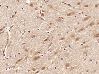
RAD26L/C9orf102 Polyclonal Antibody
Purified Rabbit Polyclonal Antibody (Pab)
- SPECIFICATION
- CITATIONS
- PROTOCOLS
- BACKGROUND

Application
| IHC-P, IHC-F, IF, ICC, E |
|---|---|
| Primary Accession | Q5T890 |
| Reactivity | Rat |
| Host | Rabbit |
| Clonality | Polyclonal |
| Calculated MW | 177 KDa |
| Physical State | Liquid |
| Immunogen | KLH conjugated synthetic peptide derived from human RAD26L/C9orf102 |
| Epitope Specificity | 461-560/1561 |
| Isotype | IgG |
| Purity | affinity purified by Protein A |
| Buffer | 0.01M TBS (pH7.4) with 1% BSA, 0.02% Proclin300 and 50% Glycerol. |
| SUBCELLULAR LOCATION | Nucleus. |
| SIMILARITY | Belongs to the SNF2/RAD54 helicase family. Contains 1 helicase ATP-binding domain. Contains 1 helicase C-terminal domain. |
| Important Note | This product as supplied is intended for research use only, not for use in human, therapeutic or diagnostic applications. |
| Background Descriptions | This gene encodes a member of the Snf2 family of helicase-like proteins. The encoded protein may play a role in DNA repair and mitochondrial function. Mutations in this gene have been associated with bone marrow failure syndrome 2. Alternatively spliced transcript variants that encode different protein isoforms have been described. [provided by RefSeq, Apr 2014] |
| Gene ID | 375748 |
|---|---|
| Other Names | DNA excision repair protein ERCC-6-like 2, 3.6.4.-, DNA repair and recombination protein RAD26-like, ERCC6L2, C9orf102, RAD26L |
| Dilution | IHC-P=1:100-500,IHC-F=1:100-500,ICC=1:100-500,IF=1:100-500,ELISA=1:5000-10000 |
| Storage | Store at -20 ℃ for one year. Avoid repeated freeze/thaw cycles. When reconstituted in sterile pH 7.4 0.01M PBS or diluent of antibody the antibody is stable for at least two weeks at 2-4 ℃. |
| Name | ERCC6L2 (HGNC:26922) |
|---|---|
| Synonyms | C9orf102, RAD26L |
| Function | Promotes double-strand break (DSB) end-joining and facilitates programmed recombination by controlling how DNA ends are joined in a spatially oriented manner during repair (By similarity). Also plays a role in DNA repair by restricting DNA end resection in double strand break (DSB) repair (PubMed:24507776, PubMed:37014751). Facilitates replication of complex DNA regions and regulates the maintenance of chromatin structure (PubMed:37014751). |
| Cellular Location | Nucleus. Cytoplasm, cytoskeleton, microtubule organizing center, centrosome Mitochondrion. Chromosome, centromere. Note=Colocalizes with NEK6 in the centrosome (PubMed:20873783). In response to DNA damage, translocates from the cytosol to mitochondria and nucleus in a reactive oxygen species (ROS)-dependent manner (PubMed:24507776). Centromeric localization is facilitated by its interaction with PCNA (PubMed:37014751). |
| Tissue Location | Expressed in bone marrow (at protein level). |
Citations (0)

Thousands of laboratories across the world have published research that depended on the performance of antibodies from Abcepta to advance their research. Check out links to articles that cite our products in major peer-reviewed journals, organized by research category.
Submit your citation using an Abcepta antibody to
info@abcepta.com, and receive a free "I Love Antibodies" mug.
info@abcepta.com, and receive a free "I Love Antibodies" mug.

Application Protocols
Provided below are standard protocols that you may find useful for product applications.
Abcepta welcomes feedback from its customers.
If you have used an Abcepta product and would like to share how it has performed, please click on the "Submit Review" button and provide the requested information. Our staff will examine and post your review and contact you if needed.
If you have any additional inquiries please email technical services at tech@abcepta.com.
$ 385.00
Cat# AP57639
Ordering Information
United States
AlbaniaAustraliaAustriaBelgiumBosnia & HerzegovinaBrazilBulgariaCanadaCentral AmericaChinaCroatiaCyprusCzech RepublicDenmarkEstoniaFinlandFranceGermanyGreeceHong KongHungaryIcelandIndiaIndonesiaIrelandIsraelItalyJapanLatviaLithuaniaLuxembourgMacedoniaMalaysiaMaltaMexicoNetherlandsNew ZealandNorwayPakistanPolandPortugalRomaniaSerbiaSingaporeSlovakiaSloveniaSouth AfricaSouth KoreaSpainSwedenSwitzerlandTaiwanTurkeyUnited KingdomUnited StatesVietnamWorldwideOthers
USA Headquarters
(888) 735-7227 / (858) 622-0099 or (858) 875-1900
Other Products
Shipping Information
Domestic orders (in stock items)
Shipped out the same day. Orders placed after 1 PM (PST) will ship out the next business day.
International orders
Contact your local distributors
