


 Foundational characteristics of cancer include proliferation, angiogenesis, migration, evasion of apoptosis, and cellular immortality. Find key markers for these cellular processes and antibodies to detect them.
 The SUMOplot™ Analysis Program predicts and scores sumoylation sites in your protein. SUMOylation is a post-translational modification involved in various cellular processes, such as nuclear-cytosolic transport, transcriptional regulation, apoptosis, protein stability, response to stress, and progression through the cell cycle.
 The Autophagy Receptor Motif Plotter predicts and scores autophagy receptor binding sites in your protein. Identifying proteins connected to this pathway is critical to understanding the role of autophagy in physiological as well as pathological processes such as development, differentiation, neurodegenerative diseases, stress, infection, and cancer.
PCDHB3 Antibody (N-term)
Affinity Purified Rabbit Polyclonal Antibody (Pab)
- SPECIFICATION
- CITATIONS
- PROTOCOLS
- BACKGROUND

Application
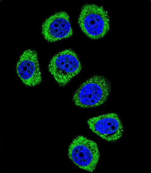
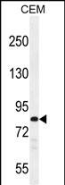
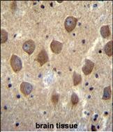
| IF, IHC-P, WB, E |
|---|---|
| Primary Accession | Q9Y5E6 |
| Other Accession | NP_061760.1 |
| Reactivity | Human |
| Host | Rabbit |
| Clonality | Polyclonal |
| Isotype | Rabbit IgG |
| Calculated MW | 86714 Da |
| Antigen Region | 12-39 aa |
| Gene ID | 56132 |
|---|---|
| Other Names | Protocadherin beta-3, PCDH-beta-3, PCDHB3 |
| Target/Specificity | This PCDHB3 antibody is generated from rabbits immunized with a KLH conjugated synthetic peptide between 12-39 amino acids from the N-terminal region of human PCDHB3. |
| Dilution | IF~~1:10~50 IHC-P~~1:10~50 WB~~1:1000 E~~Use at an assay dependent concentration. |
| Format | Purified polyclonal antibody supplied in PBS with 0.09% (W/V) sodium azide. This antibody is purified through a protein A column, followed by peptide affinity purification. |
| Storage | Maintain refrigerated at 2-8°C for up to 2 weeks. For long term storage store at -20°C in small aliquots to prevent freeze-thaw cycles. |
| Precautions | PCDHB3 Antibody (N-term) is for research use only and not for use in diagnostic or therapeutic procedures. |
| Name | PCDHB3 |
|---|---|
| Function | Potential calcium-dependent cell-adhesion protein. May be involved in the establishment and maintenance of specific neuronal connections in the brain. |
| Cellular Location | Cell membrane; Single-pass type I membrane protein |

Thousands of laboratories across the world have published research that depended on the performance of antibodies from Abcepta to advance their research. Check out links to articles that cite our products in major peer-reviewed journals, organized by research category.
info@abcepta.com, and receive a free "I Love Antibodies" mug.

Provided below are standard protocols that you may find useful for product applications.
Background
This gene is a member of the protocadherin beta gene cluster, one of three related gene clusters tandemly linked on chromosome five. The gene clusters demonstrate an unusual genomic organization similar to that of B-cell and T-cell receptor gene clusters. The beta cluster contains 16 genes and 3 pseudogenes, each encoding 6 extracellular cadherin domains and a cytoplasmic tail that deviates from others in the cadherin superfamily. The extracellular domains interact in a homophilic manner to specify differential cell-cell connections. Unlike the alpha and gamma clusters, the transcripts from these genes are made up of only one large exon, not sharing common 3' exons as expected. These neural cadherin-like cell adhesion proteins are integral plasma membrane proteins. Their specific functions are unknown but they most likely play a critical role in the establishment and function of specific cell-cell neural connections.
References
Frank, M., et al. Curr. Opin. Cell Biol. 14(5):557-562(2002)
Vanhalst, K., et al. FEBS Lett. 495 (1-2), 120-125 (2001) :
Wu, Q., et al. Genome Res. 11(3):389-404(2001)
Nollet, F., et al. J. Mol. Biol. 299(3):551-572(2000)
Yagi, T., et al. Genes Dev. 14(10):1169-1180(2000)
If you have used an Abcepta product and would like to share how it has performed, please click on the "Submit Review" button and provide the requested information. Our staff will examine and post your review and contact you if needed.
If you have any additional inquiries please email technical services at tech@abcepta.com.
Ordering Information
Other Products
Shipping Information
