


 Foundational characteristics of cancer include proliferation, angiogenesis, migration, evasion of apoptosis, and cellular immortality. Find key markers for these cellular processes and antibodies to detect them.
 The SUMOplot™ Analysis Program predicts and scores sumoylation sites in your protein. SUMOylation is a post-translational modification involved in various cellular processes, such as nuclear-cytosolic transport, transcriptional regulation, apoptosis, protein stability, response to stress, and progression through the cell cycle.
 The Autophagy Receptor Motif Plotter predicts and scores autophagy receptor binding sites in your protein. Identifying proteins connected to this pathway is critical to understanding the role of autophagy in physiological as well as pathological processes such as development, differentiation, neurodegenerative diseases, stress, infection, and cancer.
FOXE3 Antibody (Center)
Affinity Purified Rabbit Polyclonal Antibody (Pab)
- SPECIFICATION
- CITATIONS
- PROTOCOLS
- BACKGROUND

Application
| WB, E |
|---|---|
| Primary Accession | Q13461 |
| Other Accession | Q63250, Q9QY14, NP_036318.1 |
| Reactivity | Human |
| Predicted | Mouse, Rat |
| Host | Rabbit |
| Clonality | Polyclonal |
| Isotype | Rabbit IgG |
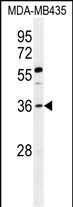
| Calculated MW | 33234 Da |
| Antigen Region | 87-114 aa |
| Gene ID | 2301 |
|---|---|
| Other Names | Forkhead box protein E3, Forkhead-related protein FKHL12, Forkhead-related transcription factor 8, FREAC-8, FOXE3, FKHL12, FREAC8 |
| Target/Specificity | This FOXE3 antibody is generated from rabbits immunized with a KLH conjugated synthetic peptide between 87-114 amino acids from the Central region of human FOXE3. |
| Dilution | WB~~1:1000 E~~Use at an assay dependent concentration. |
| Format | Purified polyclonal antibody supplied in PBS with 0.09% (W/V) sodium azide. This antibody is purified through a protein A column, followed by peptide affinity purification. |
| Storage | Maintain refrigerated at 2-8°C for up to 2 weeks. For long term storage store at -20°C in small aliquots to prevent freeze-thaw cycles. |
| Precautions | FOXE3 Antibody (Center) is for research use only and not for use in diagnostic or therapeutic procedures. |
| Name | FOXE3 |
|---|---|
| Synonyms | FKHL12, FREAC8 |
| Function | Transcription factor that controls lens epithelial cell growth through regulation of proliferation, apoptosis and cell cycle (PubMed:22527307, PubMed:25504734). During lens development, controls the ratio of the lens fiber cells to the cells of the anterior lens epithelium by regulating the rate of proliferation and differentiation (By similarity). Controls lens vesicle closure and subsequent separation of the lens vesicle from ectoderm (By similarity). Controls the expression of DNAJB1 in a pathway that is crucial for the development of the anterior segment of the eye (PubMed:27218149). |
| Cellular Location | Nucleus. |

Thousands of laboratories across the world have published research that depended on the performance of antibodies from Abcepta to advance their research. Check out links to articles that cite our products in major peer-reviewed journals, organized by research category.
info@abcepta.com, and receive a free "I Love Antibodies" mug.

Provided below are standard protocols that you may find useful for product applications.
Background
This intronless gene belongs to the forkhead family of transcription factors, which is characterized by a distinct forkhead domain. The protein encoded functions as a lens-specific transcription factor and plays an important role in vertebrate lens formation. Mutations in this gene are associated with anterior segment mesenchymal dysgenesis and congenital primary aphakia.
References
Reis, L.M., et al. Am. J. Med. Genet. A 152A (3), 582-590 (2010) :
Bremond-Gignac, D., et al. Mol. Vis. 16, 1705-1711 (2010) :
Ali, M., et al. Mol. Vis. 16, 1162-1168 (2010) :
Anjum, I., et al. Mol. Vis. 16, 549-555 (2010) :
Iseri, S.U., et al. Hum. Mutat. 30(10):1378-1386(2009)
If you have used an Abcepta product and would like to share how it has performed, please click on the "Submit Review" button and provide the requested information. Our staff will examine and post your review and contact you if needed.
If you have any additional inquiries please email technical services at tech@abcepta.com.
Ordering Information
Other Products
Shipping Information
