


 Foundational characteristics of cancer include proliferation, angiogenesis, migration, evasion of apoptosis, and cellular immortality. Find key markers for these cellular processes and antibodies to detect them.
 The SUMOplot™ Analysis Program predicts and scores sumoylation sites in your protein. SUMOylation is a post-translational modification involved in various cellular processes, such as nuclear-cytosolic transport, transcriptional regulation, apoptosis, protein stability, response to stress, and progression through the cell cycle.
 The Autophagy Receptor Motif Plotter predicts and scores autophagy receptor binding sites in your protein. Identifying proteins connected to this pathway is critical to understanding the role of autophagy in physiological as well as pathological processes such as development, differentiation, neurodegenerative diseases, stress, infection, and cancer.
MAD1L1 Antibody (N-term)
Affinity Purified Rabbit Polyclonal Antibody (Pab)
- SPECIFICATION
- CITATIONS: 1
- PROTOCOLS
- BACKGROUND

Application
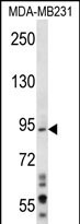
| WB, E |
|---|---|
| Primary Accession | Q9Y6D9 |
| Other Accession | NP_001013859.1, NP_001013858.1, NP_003541.2 |
| Reactivity | Human |
| Host | Rabbit |
| Clonality | Polyclonal |
| Isotype | Rabbit IgG |
| Calculated MW | 83067 Da |
| Antigen Region | 124-151 aa |
| Gene ID | 8379 |
|---|---|
| Other Names | Mitotic spindle assembly checkpoint protein MAD1, Mitotic arrest deficient 1-like protein 1, MAD1-like protein 1, Mitotic checkpoint MAD1 protein homolog, HsMAD1, hMAD1, Tax-binding protein 181, MAD1L1, MAD1, TXBP181 |
| Target/Specificity | This MAD1L1 antibody is generated from rabbits immunized with a KLH conjugated synthetic peptide between 124-151 amino acids from the N-terminal region of human MAD1L1. |
| Dilution | WB~~1:1000 E~~Use at an assay dependent concentration. |
| Format | Purified polyclonal antibody supplied in PBS with 0.09% (W/V) sodium azide. This antibody is purified through a protein A column, followed by peptide affinity purification. |
| Storage | Maintain refrigerated at 2-8°C for up to 2 weeks. For long term storage store at -20°C in small aliquots to prevent freeze-thaw cycles. |
| Precautions | MAD1L1 Antibody (N-term) is for research use only and not for use in diagnostic or therapeutic procedures. |
| Name | MAD1L1 |
|---|---|
| Synonyms | MAD1, TXBP181 |
| Function | Component of the spindle-assembly checkpoint that prevents the onset of anaphase until all chromosomes are properly aligned at the metaphase plate (PubMed:10049595, PubMed:20133940, PubMed:29162720). Forms a heterotetrameric complex with the closed conformation form of MAD2L1 (C-MAD2) at unattached kinetochores during prometaphase, recruits an open conformation of MAD2L1 (O-MAD2) and promotes the conversion of O-MAD2 to C-MAD2, which ensures mitotic checkpoint signaling (PubMed:29162720). |
| Cellular Location | Nucleus. Chromosome, centromere, kinetochore. Nucleus envelope Cytoplasm, cytoskeleton, microtubule organizing center, centrosome. Cytoplasm, cytoskeleton, spindle. Cytoplasm, cytoskeleton, spindle pole. Note=Co- localizes with TPR at the nucleus envelope during interphase and throughout the cell cycle (PubMed:18981471, PubMed:22351768). From the beginning to the end of mitosis, it is seen to move from a diffusely nuclear distribution to the centrosome, to the spindle midzone and finally to the midbody (PubMed:9546394). Localizes to kinetochores during prometaphase (PubMed:22351768, PubMed:29162720). Does not localize to kinetochores during metaphase (PubMed:29162720) Colocalizes with NEK2 at the kinetochore (PubMed:14978040). Colocalizes with IK at spindle poles during metaphase and anaphase (PubMed:22351768). |
| Tissue Location | [Isoform 1]: Expressed in hepatocellular carcinomas and hepatoma cell lines (at protein level) |

Provided below are standard protocols that you may find useful for product applications.
Background
MAD1L1 is a component of the mitotic spindle-assembly checkpoint that prevents the onset of anaphase until all chromosome are properly aligned at the metaphase plate. MAD1L1 functions as a homodimer and interacts with MAD2L1. MAD1L1 may play a role in cell cycle control and tumor suppression. Three transcript variants encoding the same protein have been found for this gene. [provided by RefSeq].
References
Shimada, M., et al. Hum. Genet. 128(4):433-441(2010)
Guo, Y., et al. J. Med. Genet. 47(9):616-622(2010)
Wang, H.B., et al. J. Gastrointest. Surg. 14(8):1227-1234(2010)
Hewitt, L., et al. J. Cell Biol. 190(1):25-34(2010)
Ge, Z., et al. FASEB J. 24(2):579-586(2010)
If you have used an Abcepta product and would like to share how it has performed, please click on the "Submit Review" button and provide the requested information. Our staff will examine and post your review and contact you if needed.
If you have any additional inquiries please email technical services at tech@abcepta.com.
Ordering Information
Other Products
Shipping Information
