


 Foundational characteristics of cancer include proliferation, angiogenesis, migration, evasion of apoptosis, and cellular immortality. Find key markers for these cellular processes and antibodies to detect them.
 The SUMOplot™ Analysis Program predicts and scores sumoylation sites in your protein. SUMOylation is a post-translational modification involved in various cellular processes, such as nuclear-cytosolic transport, transcriptional regulation, apoptosis, protein stability, response to stress, and progression through the cell cycle.
 The Autophagy Receptor Motif Plotter predicts and scores autophagy receptor binding sites in your protein. Identifying proteins connected to this pathway is critical to understanding the role of autophagy in physiological as well as pathological processes such as development, differentiation, neurodegenerative diseases, stress, infection, and cancer.
DTNA Antibody (C-term)
Affinity Purified Rabbit Polyclonal Antibody (Pab)
- SPECIFICATION
- CITATIONS
- PROTOCOLS
- BACKGROUND

Application
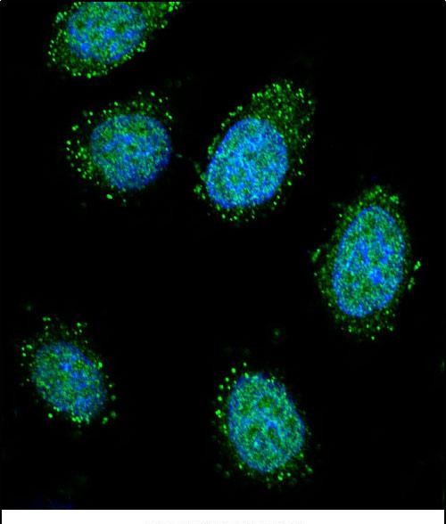
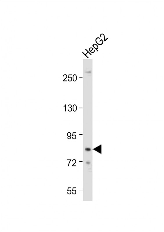
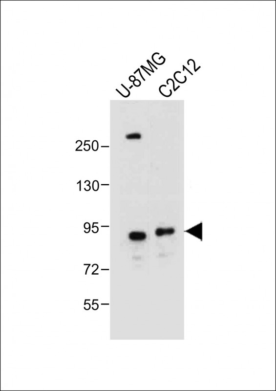
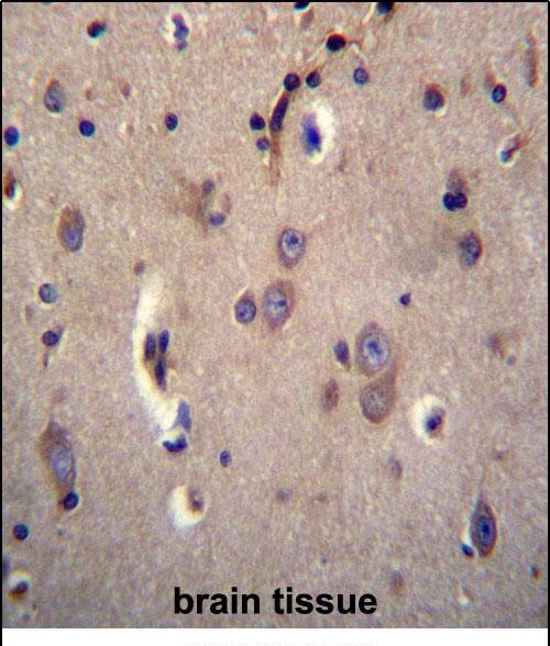
| IF, IHC-P, WB, E |
|---|---|
| Primary Accession | Q9Y4J8 |
| Other Accession | NP_116760.2, NP_001383.2 |
| Reactivity | Human, Mouse |
| Host | Rabbit |
| Clonality | Polyclonal |
| Isotype | Rabbit IgG |
| Calculated MW | 83901 Da |
| Antigen Region | 692-721 aa |
| Gene ID | 1837 |
|---|---|
| Other Names | Dystrobrevin alpha, DTN-A, Alpha-dystrobrevin, Dystrophin-related protein 3, DTNA, DRP3 |
| Target/Specificity | This DTNA antibody is generated from rabbits immunized with a KLH conjugated synthetic peptide between 692-721 amino acids from the C-terminal region of human DTNA. |
| Dilution | IF~~1:10~50 IHC-P~~1:10~50 WB~~1:1000 E~~Use at an assay dependent concentration. |
| Format | Purified polyclonal antibody supplied in PBS with 0.09% (W/V) sodium azide. This antibody is purified through a protein A column, followed by peptide affinity purification. |
| Storage | Maintain refrigerated at 2-8°C for up to 2 weeks. For long term storage store at -20°C in small aliquots to prevent freeze-thaw cycles. |
| Precautions | DTNA Antibody (C-term) is for research use only and not for use in diagnostic or therapeutic procedures. |
| Name | DTNA (HGNC:3057) |
|---|---|
| Synonyms | DRP3 |
| Function | May be involved in the formation and stability of synapses as well as being involved in the clustering of nicotinic acetylcholine receptors. |
| Cellular Location | Cytoplasm. Synapse. Cell membrane. Note=In peripheral nerves, colocalizes with MAGEE1 in the Schwann cell membrane. |
| Tissue Location | Highly expressed in brain, skeletal and cardiac muscles, and expressed at lower levels in lung, liver and pancreas Isoform 2 is not expressed in cardiac muscle. Isoform 7 and isoform 8 are only expressed in muscle |

Thousands of laboratories across the world have published research that depended on the performance of antibodies from Abcepta to advance their research. Check out links to articles that cite our products in major peer-reviewed journals, organized by research category.
info@abcepta.com, and receive a free "I Love Antibodies" mug.

Provided below are standard protocols that you may find useful for product applications.
Background
The protein encoded by this gene belongs to the dystrobrevin subfamily of the dystrophin family. This protein is a component of the dystrophin-associated protein complex (DPC), which consists of dystrophin and several integral and peripheral membrane proteins, including dystroglycans, sarcoglycans, syntrophins and alpha- and beta-dystrobrevin. The DPC localizes to the sarcolemma and its disruption is associated with various forms of muscular dystrophy. Mutations in this gene are associated with left ventricular noncompaction with congenital heart defects. Multiple alternatively spliced transcript variants encoding different isoforms have been identified for this gene.
References
Rose, J.E., et al. Mol. Med. 16 (7-8), 247-253 (2010) :
Bohm, S.V., et al. BMC Biol. 7, 85 (2009) :
Nakamori, M., et al. Neurology 70(9):677-685(2008)
Lamesch, P., et al. Genomics 89(3):307-315(2007)
Lim, J., et al. Cell 125(4):801-814(2006)
If you have used an Abcepta product and would like to share how it has performed, please click on the "Submit Review" button and provide the requested information. Our staff will examine and post your review and contact you if needed.
If you have any additional inquiries please email technical services at tech@abcepta.com.
Ordering Information
Other Products
Shipping Information
