


 Foundational characteristics of cancer include proliferation, angiogenesis, migration, evasion of apoptosis, and cellular immortality. Find key markers for these cellular processes and antibodies to detect them.
 The SUMOplot™ Analysis Program predicts and scores sumoylation sites in your protein. SUMOylation is a post-translational modification involved in various cellular processes, such as nuclear-cytosolic transport, transcriptional regulation, apoptosis, protein stability, response to stress, and progression through the cell cycle.
 The Autophagy Receptor Motif Plotter predicts and scores autophagy receptor binding sites in your protein. Identifying proteins connected to this pathway is critical to understanding the role of autophagy in physiological as well as pathological processes such as development, differentiation, neurodegenerative diseases, stress, infection, and cancer.
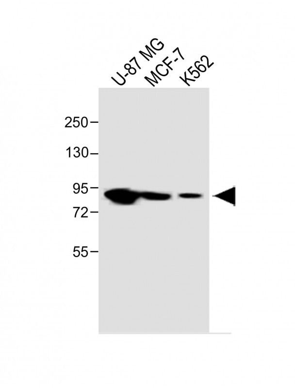
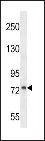
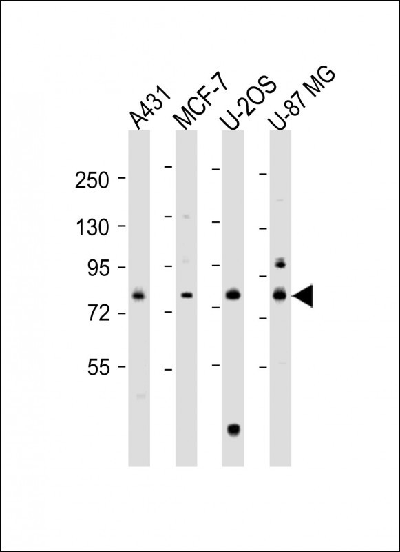
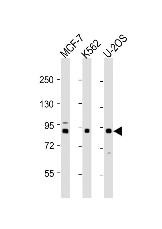
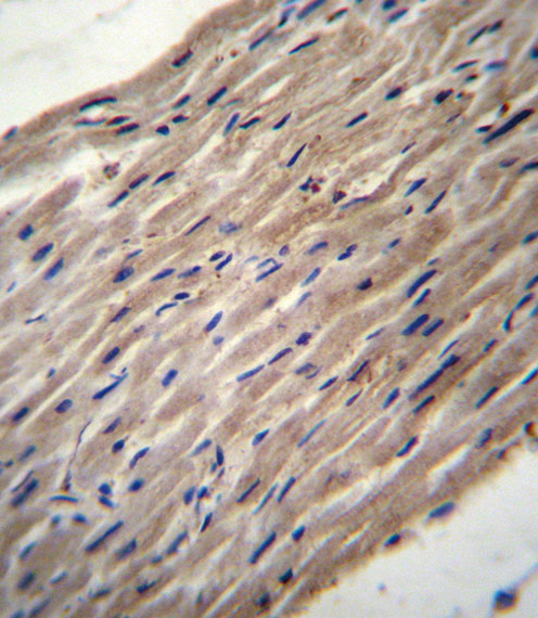
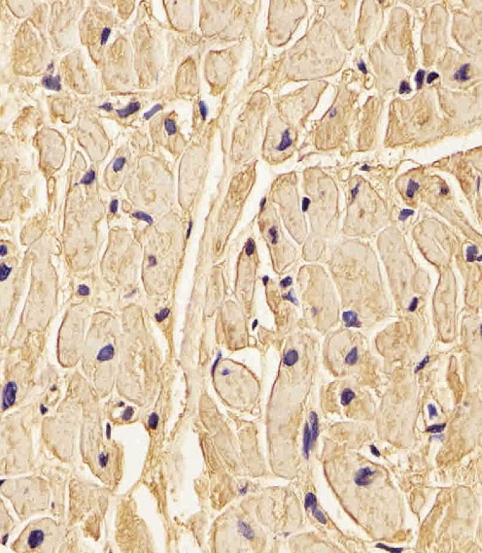
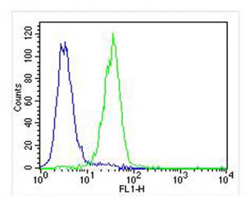
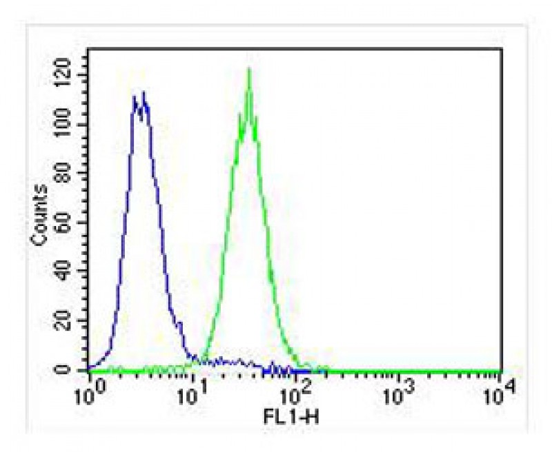
PLOD1 Antibody (N-term)
Affinity Purified Rabbit Polyclonal Antibody (Pab)
- SPECIFICATION
- CITATIONS: 3
- PROTOCOLS
- BACKGROUND

Application
| WB, IHC-P, FC, E |
|---|---|
| Primary Accession | Q02809 |
| Other Accession | Q9R0E2, NP_000293.2 |
| Reactivity | Human |
| Predicted | Mouse |
| Host | Rabbit |
| Clonality | Polyclonal |
| Isotype | Rabbit IgG |
| Calculated MW | 83550 Da |
| Antigen Region | 66-94 aa |
| Gene ID | 5351 |
|---|---|
| Other Names | Procollagen-lysine, 2-oxoglutarate 5-dioxygenase 1, Lysyl hydroxylase 1, LH1, PLOD1, LLH, PLOD |
| Target/Specificity | This PLOD1 antibody is generated from rabbits immunized with a KLH conjugated synthetic peptide between 66-94 amino acids from the N-terminal region of human PLOD1. |
| Dilution | WB~~1:1000-1:2000 IHC-P~~1:25 FC~~1:25 E~~Use at an assay dependent concentration. |
| Format | Purified polyclonal antibody supplied in PBS with 0.09% (W/V) sodium azide. This antibody is purified through a protein A column, followed by peptide affinity purification. |
| Storage | Maintain refrigerated at 2-8°C for up to 2 weeks. For long term storage store at -20°C in small aliquots to prevent freeze-thaw cycles. |
| Precautions | PLOD1 Antibody (N-term) is for research use only and not for use in diagnostic or therapeutic procedures. |
| Name | PLOD1 |
|---|---|
| Synonyms | LLH, PLOD |
| Function | Part of a complex composed of PLOD1, P3H3 and P3H4 that catalyzes hydroxylation of lysine residues in collagen alpha chains and is required for normal assembly and cross-linkling of collagen fibrils (By similarity). Forms hydroxylysine residues in -Xaa-Lys- Gly- sequences in collagens (PubMed:10686424, PubMed:15854030, PubMed:8621606). These hydroxylysines serve as sites of attachment for carbohydrate units and are essential for the stability of the intermolecular collagen cross-links (Probable). |
| Cellular Location | Rough endoplasmic reticulum membrane; Peripheral membrane protein; Lumenal side |

Provided below are standard protocols that you may find useful for product applications.
Background
Lysyl hydroxylase is a membrane-bound homodimeric protein localized to the cisternae of the endoplasmic reticulum. The enzyme (cofactors iron and ascorbate) catalyzes the hydroxylation of lysyl residues in collagen-like peptides. The resultant hydroxylysyl groups are attachment sites for carbohydrates in collagen and thus are critical for the stability of intermolecular crosslinks. Some patients with Ehlers-Danlos syndrome type VI have deficiencies in lysyl hydroxylase activity.
References
Johnatty, S.E., et al. PLoS Genet. 6 (7), E1001016 (2010) :
Huang, Q.Y., et al. Bone 44(5):984-988(2009)
Yamada, Y., et al. Int. J. Mol. Med. 19(5):791-801(2007)
Tasker, P.N., et al. Osteoporos Int 17(7):1078-1085(2006)
Giunta, C., et al. Mol. Genet. Metab. 86 (1-2), 269-276 (2005) :
If you have used an Abcepta product and would like to share how it has performed, please click on the "Submit Review" button and provide the requested information. Our staff will examine and post your review and contact you if needed.
If you have any additional inquiries please email technical services at tech@abcepta.com.
Ordering Information
Other Products
Shipping Information
