


 Foundational characteristics of cancer include proliferation, angiogenesis, migration, evasion of apoptosis, and cellular immortality. Find key markers for these cellular processes and antibodies to detect them.
 The SUMOplot™ Analysis Program predicts and scores sumoylation sites in your protein. SUMOylation is a post-translational modification involved in various cellular processes, such as nuclear-cytosolic transport, transcriptional regulation, apoptosis, protein stability, response to stress, and progression through the cell cycle.
 The Autophagy Receptor Motif Plotter predicts and scores autophagy receptor binding sites in your protein. Identifying proteins connected to this pathway is critical to understanding the role of autophagy in physiological as well as pathological processes such as development, differentiation, neurodegenerative diseases, stress, infection, and cancer.
TNMD Antibody (Center)
Affinity Purified Rabbit Polyclonal Antibody (Pab)
- SPECIFICATION
- CITATIONS: 1
- PROTOCOLS
- BACKGROUND

Application
| WB, E |
|---|---|
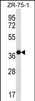
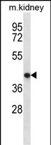
| Primary Accession | Q9H2S6 |
| Other Accession | NP_071427.2 |
| Reactivity | Human, Mouse |
| Host | Rabbit |
| Clonality | Polyclonal |
| Isotype | Rabbit IgG |
| Calculated MW | 37130 Da |
| Antigen Region | 213-241 aa |
| Gene ID | 64102 |
|---|---|
| Other Names | Tenomodulin, TeM, hTeM, Chondromodulin-1-like protein, ChM1L, hChM1L, Chondromodulin-I-like protein, Myodulin, Tendin, TNMD, CHM1L |
| Target/Specificity | This TNMD antibody is generated from rabbits immunized with a KLH conjugated synthetic peptide between 213-241 amino acids from the Central region of human TNMD. |
| Dilution | WB~~1:1000 E~~Use at an assay dependent concentration. |
| Format | Purified polyclonal antibody supplied in PBS with 0.09% (W/V) sodium azide. This antibody is purified through a protein A column, followed by peptide affinity purification. |
| Storage | Maintain refrigerated at 2-8°C for up to 2 weeks. For long term storage store at -20°C in small aliquots to prevent freeze-thaw cycles. |
| Precautions | TNMD Antibody (Center) is for research use only and not for use in diagnostic or therapeutic procedures. |
| Name | TNMD |
|---|---|
| Synonyms | CHM1L |
| Function | May be an angiogenesis inhibitor. |
| Cellular Location | [Isoform 1]: Membrane; Single-pass type II membrane protein. Nucleus envelope [Isoform 3]: Cytoplasm. |
| Tissue Location | Highly expressed in hypovascular connective tissues such as tendons. Also has strong expression in adipose tissue |

Provided below are standard protocols that you may find useful for product applications.
Background
This gene encodes a protein that is related to chondromodulin-I, which is a cartilage-specific glycoprotein that functions to stimulate chondrocyte growth and to inhibit tube formation of endothelial cells. This protein is also an angiogenesis inhibitor. Genetic variation in this gene is associated with a risk for type 2 diabetes, central obesity and serum levels of systemic immune mediators in a body size-dependent manner. This gene is also a candidate gene for age-related macular degeneration, though a direct link has yet to be demonstrated.
References
Saiki, A., et al. J. Clin. Endocrinol. Metab. 94(10):3987-3994(2009)
Tolppanen, A.M., et al. Neurobiol. Aging (2009) In press :
Tolppanen, A.M., et al. Mol. Vis. 15, 762-770 (2009) :
Tolppanen, A.M., et al. Int J Obes (Lond) 32(12):1868-1872(2008)
Kimura, N., et al. Circulation 118(17):1737-1747(2008)
If you have used an Abcepta product and would like to share how it has performed, please click on the "Submit Review" button and provide the requested information. Our staff will examine and post your review and contact you if needed.
If you have any additional inquiries please email technical services at tech@abcepta.com.
Ordering Information
Other Products
Shipping Information
