


 Foundational characteristics of cancer include proliferation, angiogenesis, migration, evasion of apoptosis, and cellular immortality. Find key markers for these cellular processes and antibodies to detect them.
 The SUMOplot™ Analysis Program predicts and scores sumoylation sites in your protein. SUMOylation is a post-translational modification involved in various cellular processes, such as nuclear-cytosolic transport, transcriptional regulation, apoptosis, protein stability, response to stress, and progression through the cell cycle.
 The Autophagy Receptor Motif Plotter predicts and scores autophagy receptor binding sites in your protein. Identifying proteins connected to this pathway is critical to understanding the role of autophagy in physiological as well as pathological processes such as development, differentiation, neurodegenerative diseases, stress, infection, and cancer.
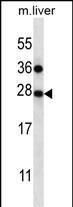
TM4SF1 Antibody (N-term)
Affinity Purified Rabbit Polyclonal Antibody (Pab)
- SPECIFICATION
- CITATIONS: 1
- PROTOCOLS
- BACKGROUND

Application
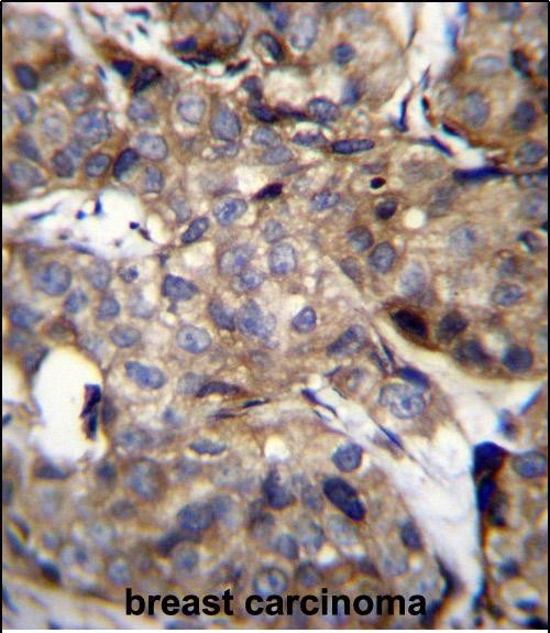
| WB, IHC-P, E |
|---|---|
| Primary Accession | P30408 |
| Other Accession | NP_055035.1 |
| Reactivity | Human, Mouse |
| Host | Rabbit |
| Clonality | Polyclonal |
| Isotype | Rabbit IgG |
| Calculated MW | 21632 Da |
| Antigen Region | 16-45 aa |
| Gene ID | 4071 |
|---|---|
| Other Names | Transmembrane 4 L6 family member 1, Membrane component chromosome 3 surface marker 1, Tumor-associated antigen L6, TM4SF1, M3S1, TAAL6 |
| Target/Specificity | This TM4SF1 antibody is generated from rabbits immunized with a KLH conjugated synthetic peptide between 16-45 amino acids from the N-terminal region of human TM4SF1. |
| Dilution | WB~~1:1000 IHC-P~~1:10~50 E~~Use at an assay dependent concentration. |
| Format | Purified polyclonal antibody supplied in PBS with 0.09% (W/V) sodium azide. This antibody is purified through a protein A column, followed by peptide affinity purification. |
| Storage | Maintain refrigerated at 2-8°C for up to 2 weeks. For long term storage store at -20°C in small aliquots to prevent freeze-thaw cycles. |
| Precautions | TM4SF1 Antibody (N-term) is for research use only and not for use in diagnostic or therapeutic procedures. |
| Name | TM4SF1 |
|---|---|
| Synonyms | M3S1, TAAL6 |
| Cellular Location | Membrane; Multi-pass membrane protein. Note=Colocalizes with SDCBP2 in the apical region of the cell (PubMed:11102519). |
| Tissue Location | Highly expressed in lung, breast, colon and ovarian carcinomas. It is also present on some normal cells, endothelial cells in particular |

Provided below are standard protocols that you may find useful for product applications.
Background
The protein encoded by this gene is a member of the transmembrane 4 superfamily, also known as the tetraspanin family. Most of these members are cell-surface proteins that are characterized by the presence of four hydrophobic domains. The proteins mediate signal transduction events that play a role in the regulation of cell development, activation, growth and motility. This encoded protein is a cell surface antigen and is highly expressed in different carcinomas.
References
Gordon, G.J., et al. J. Natl. Cancer Inst. 101(9):678-686(2009)
Lekishvili, T., et al. J. Cell. Sci. 121 (PT 5), 685-694 (2008) :
Stelzl, U., et al. Cell 122(6):957-968(2005)
Chang, Y.W., et al. Int. J. Cancer 116(2):243-252(2005)
Kao, Y.R., et al. Clin. Cancer Res. 9(7):2807-2816(2003)
If you have used an Abcepta product and would like to share how it has performed, please click on the "Submit Review" button and provide the requested information. Our staff will examine and post your review and contact you if needed.
If you have any additional inquiries please email technical services at tech@abcepta.com.
Ordering Information
Other Products
Shipping Information
