


 Foundational characteristics of cancer include proliferation, angiogenesis, migration, evasion of apoptosis, and cellular immortality. Find key markers for these cellular processes and antibodies to detect them.
 The SUMOplot™ Analysis Program predicts and scores sumoylation sites in your protein. SUMOylation is a post-translational modification involved in various cellular processes, such as nuclear-cytosolic transport, transcriptional regulation, apoptosis, protein stability, response to stress, and progression through the cell cycle.
 The Autophagy Receptor Motif Plotter predicts and scores autophagy receptor binding sites in your protein. Identifying proteins connected to this pathway is critical to understanding the role of autophagy in physiological as well as pathological processes such as development, differentiation, neurodegenerative diseases, stress, infection, and cancer.
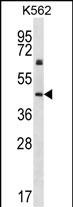
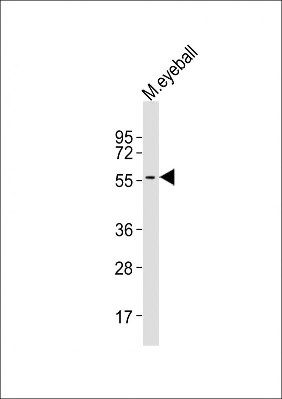
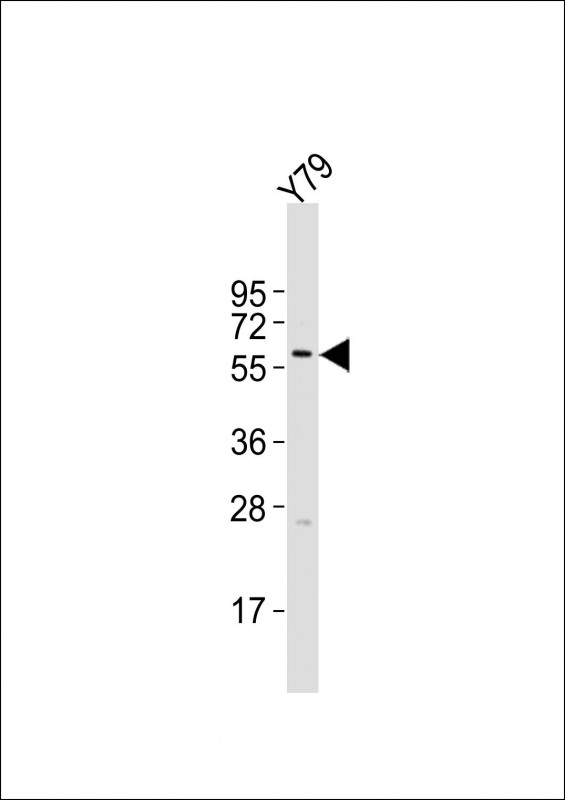
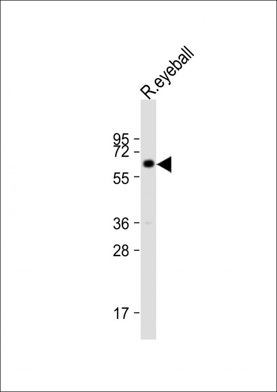
KRT12 Antibody (C-term)
Affinity Purified Rabbit Polyclonal Antibody (Pab)
- SPECIFICATION
- CITATIONS: 2
- PROTOCOLS
- BACKGROUND

Application
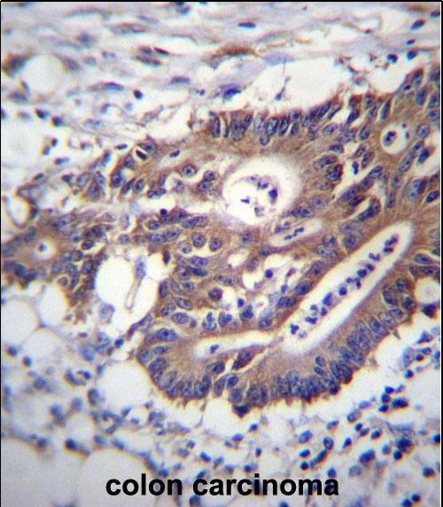
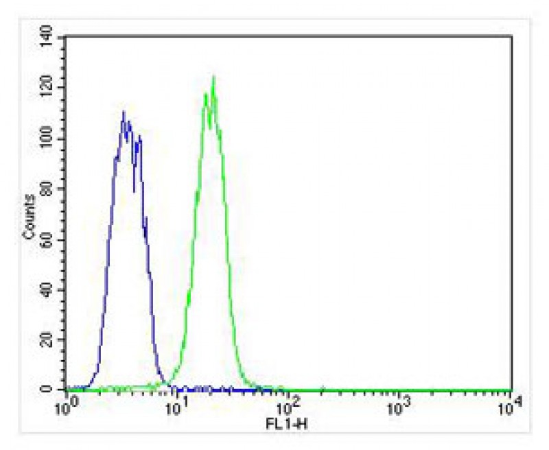
| WB, IHC-P, FC, E |
|---|---|
| Primary Accession | Q99456 |
| Other Accession | NP_000214.1 |
| Reactivity | Human, Mouse, Rat |
| Host | Rabbit |
| Clonality | Polyclonal |
| Isotype | Rabbit IgG |
| Calculated MW | 53511 Da |
| Antigen Region | 442-471 aa |
| Gene ID | 3859 |
|---|---|
| Other Names | Keratin, type I cytoskeletal 12, Cytokeratin-12, CK-12, Keratin-12, K12, KRT12 |
| Target/Specificity | This KRT12 antibody is generated from rabbits immunized with a KLH conjugated synthetic peptide between 442-471 amino acids from the C-terminal region of human KRT12. |
| Dilution | WB~~1:2000 IHC-P~~1:10~50 FC~~1:25 E~~Use at an assay dependent concentration. |
| Format | Purified polyclonal antibody supplied in PBS with 0.09% (W/V) sodium azide. This antibody is purified through a protein A column, followed by peptide affinity purification. |
| Storage | Maintain refrigerated at 2-8°C for up to 2 weeks. For long term storage store at -20°C in small aliquots to prevent freeze-thaw cycles. |
| Precautions | KRT12 Antibody (C-term) is for research use only and not for use in diagnostic or therapeutic procedures. |
| Name | KRT12 |
|---|---|
| Function | Involved in corneal epithelium organization, integrity and corneal keratin expression. |
| Tissue Location | Expressed in the corneal epithelium (at protein level). |

Provided below are standard protocols that you may find useful for product applications.
Background
KRT12 encodes the type I intermediate filament chain keratin 12, expressed in corneal epithelia. Mutations in this gene lead to Meesmann corneal dystrophy.
References
Clausen, I., et al. Mol. Vis. 16, 954-960 (2010) :
Seto, T., et al. Jpn. J. Ophthalmol. 52(3):224-226(2008)
Nielsen, K., et al. Cornea 27(1):100-102(2008)
Sullivan, L.S., et al. Mol. Vis. 13, 975-980 (2007) :
Schweizer, J., et al. J. Cell Biol. 174(2):169-174(2006)
If you have used an Abcepta product and would like to share how it has performed, please click on the "Submit Review" button and provide the requested information. Our staff will examine and post your review and contact you if needed.
If you have any additional inquiries please email technical services at tech@abcepta.com.
Ordering Information
Other Products
Shipping Information
