


 Foundational characteristics of cancer include proliferation, angiogenesis, migration, evasion of apoptosis, and cellular immortality. Find key markers for these cellular processes and antibodies to detect them.
 The SUMOplot™ Analysis Program predicts and scores sumoylation sites in your protein. SUMOylation is a post-translational modification involved in various cellular processes, such as nuclear-cytosolic transport, transcriptional regulation, apoptosis, protein stability, response to stress, and progression through the cell cycle.
 The Autophagy Receptor Motif Plotter predicts and scores autophagy receptor binding sites in your protein. Identifying proteins connected to this pathway is critical to understanding the role of autophagy in physiological as well as pathological processes such as development, differentiation, neurodegenerative diseases, stress, infection, and cancer.
SHANK2 Antibody (Center)
Affinity Purified Rabbit Polyclonal Antibody (Pab)
- SPECIFICATION
- CITATIONS
- PROTOCOLS
- BACKGROUND

Application
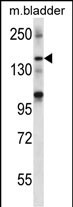
| WB, E |
|---|---|
| Primary Accession | Q9UPX8 |
| Other Accession | Q80Z38, NP_036441.2 |
| Reactivity | Mouse |
| Host | Rabbit |
| Clonality | Polyclonal |
| Isotype | Rabbit IgG |
| Calculated MW | 201261 Da |
| Antigen Region | 879-908 aa |
| Gene ID | 22941 |
|---|---|
| Other Names | SH3 and multiple ankyrin repeat domains protein 2, Shank2, Cortactin-binding protein 1, CortBP1, Proline-rich synapse-associated protein 1, SHANK2, CORTBP1, KIAA1022, PROSAP1 |
| Target/Specificity | This SHANK2 antibody is generated from rabbits immunized with a KLH conjugated synthetic peptide between 879-908 amino acids from the Central region of human SHANK2. |
| Dilution | WB~~1:1000 E~~Use at an assay dependent concentration. |
| Format | Purified polyclonal antibody supplied in PBS with 0.09% (W/V) sodium azide. This antibody is purified through a protein A column, followed by peptide affinity purification. |
| Storage | Maintain refrigerated at 2-8°C for up to 2 weeks. For long term storage store at -20°C in small aliquots to prevent freeze-thaw cycles. |
| Precautions | SHANK2 Antibody (Center) is for research use only and not for use in diagnostic or therapeutic procedures. |
| Name | SHANK2 |
|---|---|
| Synonyms | CORTBP1, KIAA1022, PROSAP1 |
| Function | Seems to be an adapter protein in the postsynaptic density (PSD) of excitatory synapses that interconnects receptors of the postsynaptic membrane including NMDA-type and metabotropic glutamate receptors, and the actin-based cytoskeleton. May play a role in the structural and functional organization of the dendritic spine and synaptic junction. |
| Cellular Location | Apical cell membrane. Cytoplasm. Synapse. Postsynaptic density. Cell projection, growth cone. Cell projection, dendritic spine. Note=Colocalizes with cortactin in growth cones in differentiating hippocampal neurons Colocalized with PDE4D to the apical membrane of colonic crypt cells (By similarity). |
| Tissue Location | Isoform 3 is present in epithelial colonic cells (at protein level). |

Thousands of laboratories across the world have published research that depended on the performance of antibodies from Abcepta to advance their research. Check out links to articles that cite our products in major peer-reviewed journals, organized by research category.
info@abcepta.com, and receive a free "I Love Antibodies" mug.

Provided below are standard protocols that you may find useful for product applications.
Background
This gene encodes a protein that is a member of the Shank family of synaptic proteins that may function as molecular scaffolds in the postsynaptic density (PSD). Shank proteins contain multiple domains for protein-protein interaction, including ankyrin repeats, an SH3 domain, a PSD-95/Dlg/ZO-1 domain, a sterile alpha motif domain, and a proline-rich region. This particular family member contains a PDZ domain, a consensus sequence for cortactin SH3 domain-binding peptides and a sterile alpha motif. The alternative splicing demonstrated in Shank genes has been suggested as a mechanism for regulating the molecular structure of Shank and the spectrum of Shank-interacting proteins in the PSDs of adult and developing brain. Two alternative splice variants, encoding distinct isoforms, are reported. Additional splice variants exist but their full-length nature has not been determined. [provided by RefSeq].
References
Pinto, D., et al. Nature 466(7304):368-372(2010)
Berkel, S., et al. Nat. Genet. 42(6):489-491(2010)
Lee, J.S., et al. J. Biol. Chem. 285(11):8104-8113(2010)
Wu, C., et al. Proteomics 7(11):1775-1785(2007)
Olsen, J.V., et al. Cell 127(3):635-648(2006)
If you have used an Abcepta product and would like to share how it has performed, please click on the "Submit Review" button and provide the requested information. Our staff will examine and post your review and contact you if needed.
If you have any additional inquiries please email technical services at tech@abcepta.com.
Ordering Information
Other Products
Shipping Information
