


 Foundational characteristics of cancer include proliferation, angiogenesis, migration, evasion of apoptosis, and cellular immortality. Find key markers for these cellular processes and antibodies to detect them.
 The SUMOplot™ Analysis Program predicts and scores sumoylation sites in your protein. SUMOylation is a post-translational modification involved in various cellular processes, such as nuclear-cytosolic transport, transcriptional regulation, apoptosis, protein stability, response to stress, and progression through the cell cycle.
 The Autophagy Receptor Motif Plotter predicts and scores autophagy receptor binding sites in your protein. Identifying proteins connected to this pathway is critical to understanding the role of autophagy in physiological as well as pathological processes such as development, differentiation, neurodegenerative diseases, stress, infection, and cancer.
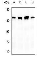
Anti-SHANK2 Antibody
Rabbit polyclonal antibody to SHANK2
- SPECIFICATION
- CITATIONS
- PROTOCOLS
- BACKGROUND

Application
| WB, IP |
|---|---|
| Primary Accession | Q9UPX8 |
| Reactivity | Human, Mouse, Rat |
| Host | Rabbit |
| Clonality | Polyclonal |
| Calculated MW | 201261 Da |
| Gene ID | 22941 |
|---|---|
| Other Names | CORTBP1; KIAA1022; PROSAP1; SH3 and multiple ankyrin repeat domains protein 2; Shank2; Cortactin-binding protein 1; CortBP1; Proline-rich synapse-associated protein 1 |
| Target/Specificity | KLH-conjugated synthetic peptide encompassing a sequence within the center region of human SHANK2. The exact sequence is proprietary. |
| Dilution | WB~~WB (1/500 - 1/1000), IP (1/10 - 1/100) IP~~N/A |
| Format | Liquid in 0.42% Potassium phosphate, 0.87% Sodium chloride, pH 7.3, 30% glycerol, and 0.09% (W/V) sodium azide. |
| Storage | Store at -20 °C.Stable for 12 months from date of receipt |
| Name | SHANK2 |
|---|---|
| Synonyms | CORTBP1, KIAA1022, PROSAP1 |
| Function | Seems to be an adapter protein in the postsynaptic density (PSD) of excitatory synapses that interconnects receptors of the postsynaptic membrane including NMDA-type and metabotropic glutamate receptors, and the actin-based cytoskeleton. May play a role in the structural and functional organization of the dendritic spine and synaptic junction. |
| Cellular Location | Apical cell membrane. Cytoplasm. Synapse. Postsynaptic density. Cell projection, growth cone. Cell projection, dendritic spine. Note=Colocalizes with cortactin in growth cones in differentiating hippocampal neurons Colocalized with PDE4D to the apical membrane of colonic crypt cells (By similarity). |
| Tissue Location | Isoform 3 is present in epithelial colonic cells (at protein level). |

Thousands of laboratories across the world have published research that depended on the performance of antibodies from Abcepta to advance their research. Check out links to articles that cite our products in major peer-reviewed journals, organized by research category.
info@abcepta.com, and receive a free "I Love Antibodies" mug.

Provided below are standard protocols that you may find useful for product applications.
Background
KLH-conjugated synthetic peptide encompassing a sequence within the center region of human SHANK2. The exact sequence is proprietary.
If you have used an Abcepta product and would like to share how it has performed, please click on the "Submit Review" button and provide the requested information. Our staff will examine and post your review and contact you if needed.
If you have any additional inquiries please email technical services at tech@abcepta.com.
Ordering Information
Other Products
Shipping Information
