


 Foundational characteristics of cancer include proliferation, angiogenesis, migration, evasion of apoptosis, and cellular immortality. Find key markers for these cellular processes and antibodies to detect them.
 The SUMOplot™ Analysis Program predicts and scores sumoylation sites in your protein. SUMOylation is a post-translational modification involved in various cellular processes, such as nuclear-cytosolic transport, transcriptional regulation, apoptosis, protein stability, response to stress, and progression through the cell cycle.
 The Autophagy Receptor Motif Plotter predicts and scores autophagy receptor binding sites in your protein. Identifying proteins connected to this pathway is critical to understanding the role of autophagy in physiological as well as pathological processes such as development, differentiation, neurodegenerative diseases, stress, infection, and cancer.
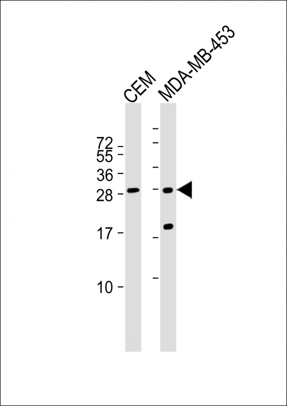
IFI6 Antibody (N-term)
Affinity Purified Rabbit Polyclonal Antibody (Pab)
- SPECIFICATION
- CITATIONS
- PROTOCOLS
- BACKGROUND

Application
| WB, E |
|---|---|
| Primary Accession | P09912-3 |
| Other Accession | NP_002029.3 |
| Reactivity | Human |
| Host | Rabbit |
| Clonality | Polyclonal |
| Isotype | Rabbit IgG |
| Antigen Region | 5-34 aa |
| Other Names | Interferon alpha-inducible protein 6, Interferon-induced protein 6-16, Ifi-6-16, IFI6, G1P3 |
|---|---|
| Target/Specificity | This IFI6 antibody is generated from rabbits immunized with a KLH conjugated synthetic peptide between 5-34 amino acids of human IFI6(P09912-3). |
| Dilution | WB~~1:1000 E~~Use at an assay dependent concentration. |
| Format | Purified polyclonal antibody supplied in PBS with 0.09% (W/V) sodium azide. This antibody is purified through a protein A column, followed by peptide affinity purification. |
| Storage | Maintain refrigerated at 2-8°C for up to 2 weeks. For long term storage store at -20°C in small aliquots to prevent freeze-thaw cycles. |
| Precautions | IFI6 Antibody (N-term) is for research use only and not for use in diagnostic or therapeutic procedures. |

Thousands of laboratories across the world have published research that depended on the performance of antibodies from Abcepta to advance their research. Check out links to articles that cite our products in major peer-reviewed journals, organized by research category.
info@abcepta.com, and receive a free "I Love Antibodies" mug.

Provided below are standard protocols that you may find useful for product applications.
Background
This gene was first identified as one of the many genes induced by interferon. The encoded protein may play a critical role in the regulation of apoptosis. A minisatellite that consists of 26 repeats of a 12 nucleotide repeating element resembling the mammalian splice donor consensus sequence begins near the end of the second exon. Alternatively spliced transcript variants that encode different isoforms by using the two downstream repeat units as splice donor sites have been described.
References
Davila, S., et al. Genes Immun. 11(3):232-238(2010)
Szegedi, K., et al. Exp. Dermatol. 19(3):269-278(2010)
Johnatty, S.E., et al. PLoS Genet. 6 (7), E1001016 (2010) :
Zhao, D., et al. Virol. J. 5, 114 (2008) :
Cheriyath, V., et al. J. Clin. Invest. 117(10):3107-3117(2007)
If you have used an Abcepta product and would like to share how it has performed, please click on the "Submit Review" button and provide the requested information. Our staff will examine and post your review and contact you if needed.
If you have any additional inquiries please email technical services at tech@abcepta.com.
Ordering Information
Other Products
Shipping Information
