


 Foundational characteristics of cancer include proliferation, angiogenesis, migration, evasion of apoptosis, and cellular immortality. Find key markers for these cellular processes and antibodies to detect them.
 The SUMOplot™ Analysis Program predicts and scores sumoylation sites in your protein. SUMOylation is a post-translational modification involved in various cellular processes, such as nuclear-cytosolic transport, transcriptional regulation, apoptosis, protein stability, response to stress, and progression through the cell cycle.
 The Autophagy Receptor Motif Plotter predicts and scores autophagy receptor binding sites in your protein. Identifying proteins connected to this pathway is critical to understanding the role of autophagy in physiological as well as pathological processes such as development, differentiation, neurodegenerative diseases, stress, infection, and cancer.
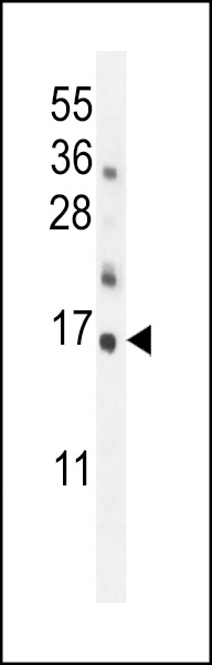
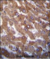
Hrk BH3 Domain Antibody
Affinity Purified Rabbit Polyclonal Antibody (Pab)
- SPECIFICATION
- CITATIONS: 1
- PROTOCOLS
- BACKGROUND

Application
| IHC-P, WB, E |
|---|---|
| Primary Accession | O00198 |
| Other Accession | NP_003797 |
| Reactivity | Human, Mouse |
| Host | Rabbit |
| Clonality | Polyclonal |
| Isotype | Rabbit IgG |
| Calculated MW | 9884 Da |
| Antigen Region | 15-50 aa |
| Gene ID | 8739 |
|---|---|
| Other Names | Activator of apoptosis harakiri, BH3-interacting domain-containing protein 3, Neuronal death protein DP5, HRK, BID3 |
| Target/Specificity | This Hrk BH3 Domain antibody is generated from rabbits immunized with a KLH conjugated synthetic peptide between 15-50 amino acids from human Hrk BH3 Domain. |
| Dilution | IHC-P~~1:10~50 WB~~1:1000 E~~Use at an assay dependent concentration. |
| Format | Purified polyclonal antibody supplied in PBS with 0.09% (W/V) sodium azide. This antibody is purified through a protein A column, followed by peptide affinity purification. |
| Storage | Maintain refrigerated at 2-8°C for up to 2 weeks. For long term storage store at -20°C in small aliquots to prevent freeze-thaw cycles. |
| Precautions | Hrk BH3 Domain Antibody is for research use only and not for use in diagnostic or therapeutic procedures. |
| Name | HRK |
|---|---|
| Synonyms | BID3 |
| Function | Promotes apoptosis. |
| Cellular Location | Membrane; Single-pass membrane protein. Mitochondrion |

Provided below are standard protocols that you may find useful for product applications.
Background
Activator of apoptosis Hrk regulates apoptosis through interaction with death-repressor proteins Bcl-2 and Bcl-X(L). The HRK protein lacks significant homology to other BCL2 family members except for an 8-amino acid region that was similar to the BCL2 homology domain-3 (BH3) motif of BIK. HRK interacts with BCL2 and BCLXL via the BH3 domain, but not with the death-promoting BCL2-related proteins BAX, BAK, or BCLXS. HRK localizes to membranes of intracellular organelles in a pattern similar to that previously reported for BCL2 and BCLXL.
References
Wakabayashi, T., et al., Neurosci. Lett. 318(2):77-80 (2002).
Inohara, N., et al., EMBO J. 16(7):1686-1694 (1997).
If you have used an Abcepta product and would like to share how it has performed, please click on the "Submit Review" button and provide the requested information. Our staff will examine and post your review and contact you if needed.
If you have any additional inquiries please email technical services at tech@abcepta.com.
Ordering Information
Other Products
Shipping Information
