


 Foundational characteristics of cancer include proliferation, angiogenesis, migration, evasion of apoptosis, and cellular immortality. Find key markers for these cellular processes and antibodies to detect them.
 The SUMOplot™ Analysis Program predicts and scores sumoylation sites in your protein. SUMOylation is a post-translational modification involved in various cellular processes, such as nuclear-cytosolic transport, transcriptional regulation, apoptosis, protein stability, response to stress, and progression through the cell cycle.
 The Autophagy Receptor Motif Plotter predicts and scores autophagy receptor binding sites in your protein. Identifying proteins connected to this pathway is critical to understanding the role of autophagy in physiological as well as pathological processes such as development, differentiation, neurodegenerative diseases, stress, infection, and cancer.
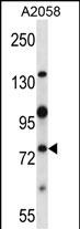
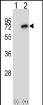
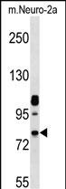
Mouse Nek8 Antibody (C-term)
Affinity Purified Rabbit Polyclonal Antibody (Pab)
- SPECIFICATION
- CITATIONS
- PROTOCOLS
- BACKGROUND

Application
| WB, E |
|---|---|
| Primary Accession | Q91ZR4 |
| Other Accession | NP_543125.1 |
| Reactivity | Human, Mouse |
| Host | Rabbit |
| Clonality | Polyclonal |
| Isotype | Rabbit IgG |
| Calculated MW | 75265 Da |
| Antigen Region | 519-547 aa |
| Gene ID | 140859 |
|---|---|
| Other Names | Serine/threonine-protein kinase Nek8, Never in mitosis A-related kinase 8, NimA-related protein kinase 8, Nek8, Jck |
| Target/Specificity | This Mouse Nek8 antibody is generated from rabbits immunized with a KLH conjugated synthetic peptide between 519-547 amino acids from the C-terminal region of mouse Nek8. |
| Dilution | WB~~1:1000 E~~Use at an assay dependent concentration. |
| Format | Purified polyclonal antibody supplied in PBS with 0.09% (W/V) sodium azide. This antibody is purified through a protein A column, followed by peptide affinity purification. |
| Storage | Maintain refrigerated at 2-8°C for up to 2 weeks. For long term storage store at -20°C in small aliquots to prevent freeze-thaw cycles. |
| Precautions | Mouse Nek8 Antibody (C-term) is for research use only and not for use in diagnostic or therapeutic procedures. |
| Name | Nek8 |
|---|---|
| Synonyms | Jck |
| Function | Required for renal tubular integrity. May regulate local cytoskeletal structure in kidney tubule epithelial cells. May regulate ciliary biogenesis through targeting of proteins to the cilia. Plays a role in organogenesis and is involved in the regulation of the Hippo signaling pathway. |
| Cellular Location | Cytoplasm. Cytoplasm, cytoskeleton. Cell projection, cilium. Cytoplasm, cytoskeleton, microtubule organizing center, centrosome {ECO:0000250|UniProtKB:Q86SG6}. Cytoplasm, cytoskeleton, cilium axoneme {ECO:0000250|UniProtKB:Q86SG6}. Note=Predominantly cytoplasmic Localizes to the proximal region of the primary cilium and is not observed in dividing cells. |
| Tissue Location | Kidney, liver, and testis. |

Thousands of laboratories across the world have published research that depended on the performance of antibodies from Abcepta to advance their research. Check out links to articles that cite our products in major peer-reviewed journals, organized by research category.
info@abcepta.com, and receive a free "I Love Antibodies" mug.

Provided below are standard protocols that you may find useful for product applications.
Background
This gene encodes a NIMA-related kinase. Members of this serine/threonine protein kinase family are structurally-related to NIMA (never in mitosis, gene A) which controls mitotic signaling in Aspergillus nidulans.
References
Hellman, N.E., et al. Proc. Natl. Acad. Sci. U.S.A. 107(43):18499-18504(2010)
Ahmadie, R., et al. J. Nutr. 140(8):1438-1444(2010)
Natoli, T.A., et al. Nat. Med. 16(7):788-792(2010)
Shiba, D., et al. Cytoskeleton (Hoboken) 67(2):112-119(2010)
Sohara, E., et al. J. Am. Soc. Nephrol. 19(3):469-476(2008)
If you have used an Abcepta product and would like to share how it has performed, please click on the "Submit Review" button and provide the requested information. Our staff will examine and post your review and contact you if needed.
If you have any additional inquiries please email technical services at tech@abcepta.com.
Ordering Information
Other Products
Shipping Information
