


 Foundational characteristics of cancer include proliferation, angiogenesis, migration, evasion of apoptosis, and cellular immortality. Find key markers for these cellular processes and antibodies to detect them.
 The SUMOplot™ Analysis Program predicts and scores sumoylation sites in your protein. SUMOylation is a post-translational modification involved in various cellular processes, such as nuclear-cytosolic transport, transcriptional regulation, apoptosis, protein stability, response to stress, and progression through the cell cycle.
 The Autophagy Receptor Motif Plotter predicts and scores autophagy receptor binding sites in your protein. Identifying proteins connected to this pathway is critical to understanding the role of autophagy in physiological as well as pathological processes such as development, differentiation, neurodegenerative diseases, stress, infection, and cancer.
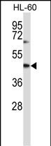
CYTH1 Antibody (N-term)
Affinity Purified Rabbit Polyclonal Antibody (Pab)
- SPECIFICATION
- CITATIONS
- PROTOCOLS
- BACKGROUND

Application
| WB, E |
|---|---|
| Primary Accession | Q15438 |
| Other Accession | P97694, Q9QX11, NP_004753.1, NP_059430.2 |
| Reactivity | Human |
| Predicted | Mouse, Rat |
| Host | Rabbit |
| Clonality | Polyclonal |
| Isotype | Rabbit IgG |
| Calculated MW | 46413 Da |
| Antigen Region | 37-65 aa |
| Gene ID | 9267 |
|---|---|
| Other Names | Cytohesin-1, PH, SEC7 and coiled-coil domain-containing protein 1, SEC7 homolog B2-1, CYTH1, D17S811E, PSCD1 |
| Target/Specificity | This CYTH1 antibody is generated from rabbits immunized with a KLH conjugated synthetic peptide between 37-65 amino acids from the N-terminal region of human CYTH1. |
| Dilution | WB~~1:1000 E~~Use at an assay dependent concentration. |
| Format | Purified polyclonal antibody supplied in PBS with 0.09% (W/V) sodium azide. This antibody is purified through a protein A column, followed by peptide affinity purification. |
| Storage | Maintain refrigerated at 2-8°C for up to 2 weeks. For long term storage store at -20°C in small aliquots to prevent freeze-thaw cycles. |
| Precautions | CYTH1 Antibody (N-term) is for research use only and not for use in diagnostic or therapeutic procedures. |
| Name | CYTH1 (HGNC:9501) |
|---|---|
| Synonyms | D17S811E, PSCD1 |
| Function | Promotes guanine-nucleotide exchange on ARF1, ARF5 and ARF6. Promotes the activation of ARF factors through replacement of GDP with GTP. Plays an important role in membrane trafficking, during junctional remodeling and epithelial polarization, through regulation of ARF6 activity. |
| Cellular Location | Cell membrane; Peripheral membrane protein. Cytoplasm, cytosol {ECO:0000250|UniProtKB:Q9QX11}. Cell junction, tight junction {ECO:0000250|UniProtKB:Q9QX11}. Cell junction, adherens junction {ECO:0000250|UniProtKB:Q9QX11}. Note=Colocalized with TJP1 during epithelial polarization. {ECO:0000250|UniProtKB:Q9QX11} |
| Tissue Location | Ubiquitous. |

Thousands of laboratories across the world have published research that depended on the performance of antibodies from Abcepta to advance their research. Check out links to articles that cite our products in major peer-reviewed journals, organized by research category.
info@abcepta.com, and receive a free "I Love Antibodies" mug.

Provided below are standard protocols that you may find useful for product applications.
Background
The protein encoded by this gene is a member of the PSCD family. Members of this family have identical structural organization that consists of an N-terminal coiled-coil motif, a central Sec7 domain, and a C-terminal pleckstrin homology (PH) domain. The coiled-coil motif is involved in homodimerization, the Sec7 domain contains guanine-nucleotide exchange protein (GEP) activity, and the PH domain interacts with phospholipids and is responsible for association of PSCDs with membranes. Members of this family appear to mediate the regulation of protein sorting and membrane trafficking. This gene is highly expressed in natural killer and peripheral T cells, and regulates the adhesiveness of integrins at the plasma membrane of lymphocytes. The encoded protein is 83% homologous to that of CYTH2.
References
El Azreq, M.A., et al. J. Immunol. 184(2):637-649(2010)
Quast, T., et al. Blood 113(23):5801-5810(2009)
Sendide, K., et al. J. Immunol. 174(7):4210-4219(2005)
Boehm, T., et al. EMBO J. 22(5):1014-1024(2003)
Mansour, M., et al. J. Biol. Chem. 277(35):32302-32309(2002)
If you have used an Abcepta product and would like to share how it has performed, please click on the "Submit Review" button and provide the requested information. Our staff will examine and post your review and contact you if needed.
If you have any additional inquiries please email technical services at tech@abcepta.com.
Ordering Information
Shipping Information
