


 Foundational characteristics of cancer include proliferation, angiogenesis, migration, evasion of apoptosis, and cellular immortality. Find key markers for these cellular processes and antibodies to detect them.
 The SUMOplot™ Analysis Program predicts and scores sumoylation sites in your protein. SUMOylation is a post-translational modification involved in various cellular processes, such as nuclear-cytosolic transport, transcriptional regulation, apoptosis, protein stability, response to stress, and progression through the cell cycle.
 The Autophagy Receptor Motif Plotter predicts and scores autophagy receptor binding sites in your protein. Identifying proteins connected to this pathway is critical to understanding the role of autophagy in physiological as well as pathological processes such as development, differentiation, neurodegenerative diseases, stress, infection, and cancer.
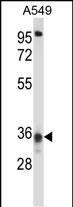
CCDC50 Antibody (Center)
Affinity Purified Rabbit Polyclonal Antibody (Pab)

- SPECIFICATION
- CITATIONS
- PROTOCOLS
- BACKGROUND

Application
| WB, IHC-P, E |
|---|---|
| Primary Accession | Q8IVM0 |
| Other Accession | Q810U0, Q810U5, NP_777568.1, NP_848018.1 |
| Reactivity | Human |
| Predicted | Mouse, Rat |
| Host | Rabbit |
| Clonality | Polyclonal |
| Isotype | Rabbit IgG |
| Calculated MW | 35822 Da |
| Antigen Region | 86-114 aa |
| Gene ID | 152137 |
|---|---|
| Other Names | Coiled-coil domain-containing protein 50, Protein Ymer, CCDC50, C3orf6 |
| Target/Specificity | This CCDC50 antibody is generated from rabbits immunized with a KLH conjugated synthetic peptide between 86-114 amino acids from the Central region of human CCDC50. |
| Dilution | WB~~1:1000 IHC-P~~1:10~50 E~~Use at an assay dependent concentration. |
| Format | Purified polyclonal antibody supplied in PBS with 0.09% (W/V) sodium azide. This antibody is purified through a protein A column, followed by peptide affinity purification. |
| Storage | Maintain refrigerated at 2-8°C for up to 2 weeks. For long term storage store at -20°C in small aliquots to prevent freeze-thaw cycles. |
| Precautions | CCDC50 Antibody (Center) is for research use only and not for use in diagnostic or therapeutic procedures. |
| Name | CCDC50 |
|---|---|
| Synonyms | C3orf6 |
| Function | Involved in EGFR signaling. |
| Cellular Location | Cytoplasm. Note=Associated with microtubules of the cytoskeleton and mitotic apparatus. |
| Tissue Location | Isoform 1 and isoform 2 are coexpressed in placenta, liver, lung, kidney and pancreas. Only isoform 1 is detected in skeletal muscle, brain and heart. |

Thousands of laboratories across the world have published research that depended on the performance of antibodies from Abcepta to advance their research. Check out links to articles that cite our products in major peer-reviewed journals, organized by research category.
info@abcepta.com, and receive a free "I Love Antibodies" mug.

Provided below are standard protocols that you may find useful for product applications.
Background
This gene encodes a soluble, cytoplasmic, tyrosine-phosphorylated protein with multiple ubiquitin-interacting domains. Mutations in this gene cause nonsyndromic, postlingual, progressive sensorineural DFNA44 hearing loss. In mouse, the protein is expressed in the inner ear during development and postnatal maturation and associates with microtubule-based structures. This protein may also function as a negative regulator of NF-kB signaling and as an effector of epidermal growth factor (EGF)-mediated cell signaling. Alternative splicing results in multiple transcript variants encoding distinct isoforms. [provided by RefSeq].
References
Farfsing, A., et al. Leukemia 23(11):2018-2026(2009)
Kameda, H., et al. Biochem. Biophys. Res. Commun. 378(4):744-749(2009)
Bohgaki, M., et al. Biochim. Biophys. Acta 1783(5):826-837(2008)
Modamio-Hoybjor, S., et al. Am. J. Hum. Genet. 80(6):1076-1089(2007)
Lamesch, P., et al. Genomics 89(3):307-315(2007)
If you have used an Abcepta product and would like to share how it has performed, please click on the "Submit Review" button and provide the requested information. Our staff will examine and post your review and contact you if needed.
If you have any additional inquiries please email technical services at tech@abcepta.com.
Ordering Information
Other Products
Shipping Information
