


 Foundational characteristics of cancer include proliferation, angiogenesis, migration, evasion of apoptosis, and cellular immortality. Find key markers for these cellular processes and antibodies to detect them.
 The SUMOplot™ Analysis Program predicts and scores sumoylation sites in your protein. SUMOylation is a post-translational modification involved in various cellular processes, such as nuclear-cytosolic transport, transcriptional regulation, apoptosis, protein stability, response to stress, and progression through the cell cycle.
 The Autophagy Receptor Motif Plotter predicts and scores autophagy receptor binding sites in your protein. Identifying proteins connected to this pathway is critical to understanding the role of autophagy in physiological as well as pathological processes such as development, differentiation, neurodegenerative diseases, stress, infection, and cancer.
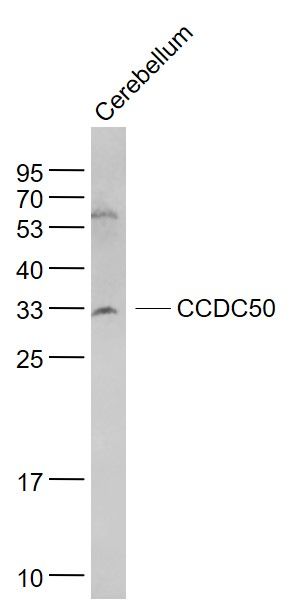
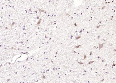
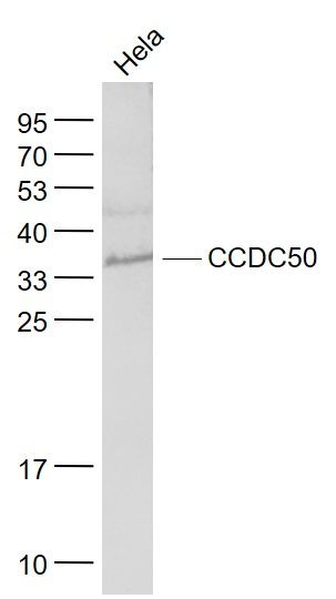
CCDC50 Polyclonal Antibody
Purified Rabbit Polyclonal Antibody (Pab)
- SPECIFICATION
- CITATIONS
- PROTOCOLS
- BACKGROUND

Application
| WB, IHC-P, IHC-F, IF, E |
|---|---|
| Primary Accession | Q8IVM0 |
| Reactivity | Rat |
| Host | Rabbit |
| Clonality | Polyclonal |
| Calculated MW | 34 KDa |
| Physical State | Liquid |
| Immunogen | KLH conjugated synthetic peptide derived from human CCDC50 |
| Epitope Specificity | 251-306/306 |
| Isotype | IgG |
| Purity | affinity purified by Protein A |
| Buffer | 0.01M TBS (pH7.4) with 1% BSA, 0.02% Proclin300 and 50% Glycerol. |
| Post-translational modifications | Phosphorylated on tyrosine residues. |
| DISEASE | Defects in CCDC50 are the cause of deafness autosomal dominant type 44 (DFNA44) . A form of non-syndromic hearing loss. It is initially moderate and affects mainly low to mid frequencies. Later, it progresses to involve all the frequencies and leads to a profound hearing loss by the 6th decade. The onset of the hearing loss occurs in the first decade of life. |
| Important Note | This product as supplied is intended for research use only, not for use in human, therapeutic or diagnostic applications. |
| Background Descriptions | This gene encodes a soluble, cytoplasmic, tyrosine-phosphorylated protein with multiple ubiquitin-interacting domains. Mutations in this gene cause nonsyndromic, postlingual, progressive sensorineural DFNA44 hearing loss. In mouse, the protein is expressed in the inner ear during development and postnatal maturation and associates with microtubule-based structures. This protein may also function as a negative regulator of NF-kB signaling and as an effector of epidermal growth factor (EGF)-mediated cell signaling. Alternative splicing results in multiple transcript variants encoding distinct isoforms. [provided by RefSeq, Oct 2008]. |
| Gene ID | 152137 |
|---|---|
| Other Names | Coiled-coil domain-containing protein 50, Protein Ymer, CCDC50, C3orf6 |
| Target/Specificity | Isoform 1 and isoform 2 are co-expressed in placenta, liver, lung, kidney and pancreas. Only isoform 1 is detected in skeletal muscle, brain and heart. |
| Dilution | WB=1:500-2000,IHC-P=1:100-500,IHC-F=1:100-500,IF=1:100-500,ELISA=1:5000-10000 |
| Storage | Store at -20 ℃ for one year. Avoid repeated freeze/thaw cycles. When reconstituted in sterile pH 7.4 0.01M PBS or diluent of antibody the antibody is stable for at least two weeks at 2-4 ℃. |
| Name | CCDC50 |
|---|---|
| Synonyms | C3orf6 |
| Function | Involved in EGFR signaling. |
| Cellular Location | Cytoplasm. Note=Associated with microtubules of the cytoskeleton and mitotic apparatus. |
| Tissue Location | Isoform 1 and isoform 2 are coexpressed in placenta, liver, lung, kidney and pancreas. Only isoform 1 is detected in skeletal muscle, brain and heart. |
Citations (0)

Thousands of laboratories across the world have published research that depended on the performance of antibodies from Abcepta to advance their research. Check out links to articles that cite our products in major peer-reviewed journals, organized by research category.
Submit your citation using an Abcepta antibody to
info@abcepta.com, and receive a free "I Love Antibodies" mug.
info@abcepta.com, and receive a free "I Love Antibodies" mug.

Application Protocols
Provided below are standard protocols that you may find useful for product applications.
Abcepta welcomes feedback from its customers.
If you have used an Abcepta product and would like to share how it has performed, please click on the "Submit Review" button and provide the requested information. Our staff will examine and post your review and contact you if needed.
If you have any additional inquiries please email technical services at tech@abcepta.com.
$ 385.00
Cat# AP58550
Ordering Information
United States
AlbaniaAustraliaAustriaBelgiumBosnia & HerzegovinaBrazilBulgariaCanadaCentral AmericaChinaCroatiaCyprusCzech RepublicDenmarkEstoniaFinlandFranceGermanyGreeceHong KongHungaryIcelandIndiaIndonesiaIrelandIsraelItalyJapanLatviaLithuaniaLuxembourgMacedoniaMalaysiaMaltaMexicoNetherlandsNew ZealandNorwayPakistanPolandPortugalRomaniaSerbiaSingaporeSlovakiaSloveniaSouth AfricaSouth KoreaSpainSwedenSwitzerlandTaiwanTurkeyUnited KingdomUnited StatesVietnamWorldwideOthers
USA Headquarters
(888) 735-7227 / (858) 622-0099 or (858) 875-1900
Other Products
Shipping Information
Domestic orders (in stock items)
Shipped out the same day. Orders placed after 1 PM (PST) will ship out the next business day.
International orders
Contact your local distributors
