


 Foundational characteristics of cancer include proliferation, angiogenesis, migration, evasion of apoptosis, and cellular immortality. Find key markers for these cellular processes and antibodies to detect them.
 The SUMOplot™ Analysis Program predicts and scores sumoylation sites in your protein. SUMOylation is a post-translational modification involved in various cellular processes, such as nuclear-cytosolic transport, transcriptional regulation, apoptosis, protein stability, response to stress, and progression through the cell cycle.
 The Autophagy Receptor Motif Plotter predicts and scores autophagy receptor binding sites in your protein. Identifying proteins connected to this pathway is critical to understanding the role of autophagy in physiological as well as pathological processes such as development, differentiation, neurodegenerative diseases, stress, infection, and cancer.
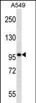
SYNPO Antibody (C-term)
Affinity Purified Rabbit Polyclonal Antibody (Pab)
- SPECIFICATION
- CITATIONS
- PROTOCOLS
- BACKGROUND

Application
| WB, E |
|---|---|
| Primary Accession | Q8N3V7 |
| Other Accession | NP_001103444.1, NP_001159681.1, NP_001159680.1 |
| Reactivity | Human |
| Host | Rabbit |
| Clonality | Polyclonal |
| Isotype | Rabbit IgG |
| Calculated MW | 99463 Da |
| Antigen Region | 654-683 aa |
| Gene ID | 11346 |
|---|---|
| Other Names | Synaptopodin, SYNPO, KIAA1029 |
| Target/Specificity | This SYNPO antibody is generated from rabbits immunized with a KLH conjugated synthetic peptide between 654-683 amino acids from the C-terminal region of human SYNPO. |
| Dilution | WB~~1:1000 E~~Use at an assay dependent concentration. |
| Format | Purified polyclonal antibody supplied in PBS with 0.09% (W/V) sodium azide. This antibody is purified through a protein A column, followed by peptide affinity purification. |
| Storage | Maintain refrigerated at 2-8°C for up to 2 weeks. For long term storage store at -20°C in small aliquots to prevent freeze-thaw cycles. |
| Precautions | SYNPO Antibody (C-term) is for research use only and not for use in diagnostic or therapeutic procedures. |
| Name | SYNPO |
|---|---|
| Synonyms | KIAA1029 |
| Function | Actin-associated protein that may play a role in modulating actin-based shape and motility of dendritic spines and renal podocyte foot processes. Seems to be essential for the formation of spine apparatuses in spines of telencephalic neurons, which is involved in synaptic plasticity (By similarity). |
| Cellular Location | Cytoplasm, cytoskeleton {ECO:0000250|UniProtKB:Q8CC35}. Cell junction, tight junction {ECO:0000250|UniProtKB:Q8CC35}. Perikaryon {ECO:0000250|UniProtKB:Q8CC35}. Cell projection, dendritic spine {ECO:0000250|UniProtKB:Q8CC35}. Postsynaptic density {ECO:0000250|UniProtKB:Q8CC35}. Synapse {ECO:0000250|UniProtKB:Q8CC35} Cytoplasm, cytosol. Note=Localized at the tight junction of cells. In brain, localized to the postsynaptic densities and in the perikarya. Associated with dendritic spines of a subset of synapses. {ECO:0000250|UniProtKB:Q8CC35} |
| Tissue Location | Expressed in cerebral cortex. |

Thousands of laboratories across the world have published research that depended on the performance of antibodies from Abcepta to advance their research. Check out links to articles that cite our products in major peer-reviewed journals, organized by research category.
info@abcepta.com, and receive a free "I Love Antibodies" mug.

Provided below are standard protocols that you may find useful for product applications.
Background
Synaptopodin is an actin-associated protein that may play a role in actin-based cell shape and motility. The name synaptopodin derives from the protein's associations with postsynaptic densities and dendritic spines and with renal podocytes (Mundel et al., 1997 [PubMed 9314539]).[supplied by OMIM].
References
Kim, E.Y., et al. Am. J. Physiol. Renal Physiol. 299 (3), F594-F604 (2010) :
Joslyn, G., et al. Alcohol. Clin. Exp. Res. 34(5):800-812(2010)
Dai, S., et al. Nephrol. Dial. Transplant. 25(3):824-835(2010)
Duning, K., et al. J. Am. Soc. Nephrol. 19(10):1891-1903(2008)
Garovic, V.D., et al. Nephrol. Dial. Transplant. 22(4):1136-1143(2007)
If you have used an Abcepta product and would like to share how it has performed, please click on the "Submit Review" button and provide the requested information. Our staff will examine and post your review and contact you if needed.
If you have any additional inquiries please email technical services at tech@abcepta.com.
Ordering Information
Other Products
Shipping Information
