


 Foundational characteristics of cancer include proliferation, angiogenesis, migration, evasion of apoptosis, and cellular immortality. Find key markers for these cellular processes and antibodies to detect them.
 The SUMOplot™ Analysis Program predicts and scores sumoylation sites in your protein. SUMOylation is a post-translational modification involved in various cellular processes, such as nuclear-cytosolic transport, transcriptional regulation, apoptosis, protein stability, response to stress, and progression through the cell cycle.
 The Autophagy Receptor Motif Plotter predicts and scores autophagy receptor binding sites in your protein. Identifying proteins connected to this pathway is critical to understanding the role of autophagy in physiological as well as pathological processes such as development, differentiation, neurodegenerative diseases, stress, infection, and cancer.
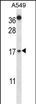
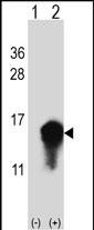
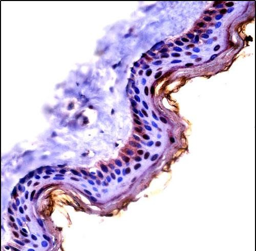
CSTA Antibody (C-term)
Affinity Purified Rabbit Polyclonal Antibody (Pab)
- SPECIFICATION
- CITATIONS
- PROTOCOLS
- BACKGROUND

Application
| IHC-P, WB, E |
|---|---|
| Primary Accession | P01040 |
| Other Accession | NP_005204.1 |
| Reactivity | Human |
| Host | Rabbit |
| Clonality | Polyclonal |
| Isotype | Rabbit IgG |
| Calculated MW | 11006 Da |
| Antigen Region | 69-98 aa |
| Gene ID | 1475 |
|---|---|
| Other Names | Cystatin-A, Cystatin-AS, Stefin-A, Cystatin-A, N-terminally processed, CSTA, STF1, STFA |
| Target/Specificity | This CSTA antibody is generated from rabbits immunized with a KLH conjugated synthetic peptide between 69-98 amino acids from the C-terminal region of human CSTA. |
| Dilution | IHC-P~~1:10~50 WB~~1:1000 E~~Use at an assay dependent concentration. |
| Format | Purified polyclonal antibody supplied in PBS with 0.09% (W/V) sodium azide. This antibody is purified through a protein A column, followed by peptide affinity purification. |
| Storage | Maintain refrigerated at 2-8°C for up to 2 weeks. For long term storage store at -20°C in small aliquots to prevent freeze-thaw cycles. |
| Precautions | CSTA Antibody (C-term) is for research use only and not for use in diagnostic or therapeutic procedures. |
| Name | CSTA |
|---|---|
| Synonyms | STF1, STFA |
| Function | This is an intracellular thiol proteinase inhibitor. Has an important role in desmosome-mediated cell-cell adhesion in the lower levels of the epidermis. |
| Cellular Location | Cytoplasm. |
| Tissue Location | Expressed in the skin throughout the epidermis. |

Thousands of laboratories across the world have published research that depended on the performance of antibodies from Abcepta to advance their research. Check out links to articles that cite our products in major peer-reviewed journals, organized by research category.
info@abcepta.com, and receive a free "I Love Antibodies" mug.

Provided below are standard protocols that you may find useful for product applications.
Background
The cystatin superfamily encompasses proteins that contain multiple cystatin-like sequences. Some of the members are active cysteine protease inhibitors, while others have lost or perhaps never acquired this inhibitory activity. There are three inhibitory families in the superfamily, including the type 1 cystatins (stefins), type 2 cystatins, and kininogens. This gene encodes a stefin that functions as a cysteine protease inhibitor, forming tight complexes with papain and the cathepsins B, H, and L. The protein is one of the precursor proteins of cornified cell envelope in keratinocytes and plays a role in epidermal development and maintenance. Stefins have been proposed as prognostic and diagnostic tools for cancer.
References
O'Seaghdha, C.M., et al. Hum. Mol. Genet. 19(21):4296-4303(2010)
Kestenbaum, B., et al. J. Am. Soc. Nephrol. 21(7):1223-1232(2010)
Takai, T., et al. J. Immunol. 183(12):7958-7965(2009)
Vasilopoulos, Y., et al. Eur. J. Hum. Genet. 16(8):1002-1009(2008)
Parker, B.S., et al. J. Pathol. 214(3):337-346(2008)
If you have used an Abcepta product and would like to share how it has performed, please click on the "Submit Review" button and provide the requested information. Our staff will examine and post your review and contact you if needed.
If you have any additional inquiries please email technical services at tech@abcepta.com.
Ordering Information
Other Products
Shipping Information
