


 Foundational characteristics of cancer include proliferation, angiogenesis, migration, evasion of apoptosis, and cellular immortality. Find key markers for these cellular processes and antibodies to detect them.
 The SUMOplot™ Analysis Program predicts and scores sumoylation sites in your protein. SUMOylation is a post-translational modification involved in various cellular processes, such as nuclear-cytosolic transport, transcriptional regulation, apoptosis, protein stability, response to stress, and progression through the cell cycle.
 The Autophagy Receptor Motif Plotter predicts and scores autophagy receptor binding sites in your protein. Identifying proteins connected to this pathway is critical to understanding the role of autophagy in physiological as well as pathological processes such as development, differentiation, neurodegenerative diseases, stress, infection, and cancer.
PPEF1 Antibody (Center)
Affinity Purified Rabbit Polyclonal Antibody (Pab)
- SPECIFICATION
- CITATIONS
- PROTOCOLS
- BACKGROUND

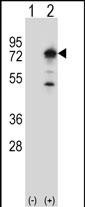
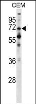
Application
| WB, E |
|---|---|
| Primary Accession | O14829 |
| Other Accession | NP_006231.2, NP_689410.1 |
| Reactivity | Human |
| Host | Rabbit |
| Clonality | Polyclonal |
| Isotype | Rabbit IgG |
| Calculated MW | 75792 Da |
| Antigen Region | 298-327 aa |
| Gene ID | 5475 |
|---|---|
| Other Names | Serine/threonine-protein phosphatase with EF-hands 1, PPEF-1, Protein phosphatase with EF calcium-binding domain, PPEF, Serine/threonine-protein phosphatase 7, PP7, PPEF1, PPEF, PPP7C |
| Target/Specificity | This PPEF1 antibody is generated from rabbits immunized with a KLH conjugated synthetic peptide between 298-327 amino acids from the Central region of human PPEF1. |
| Dilution | WB~~1:1000 E~~Use at an assay dependent concentration. |
| Format | Purified polyclonal antibody supplied in PBS with 0.09% (W/V) sodium azide. This antibody is purified through a protein A column, followed by peptide affinity purification. |
| Storage | Maintain refrigerated at 2-8°C for up to 2 weeks. For long term storage store at -20°C in small aliquots to prevent freeze-thaw cycles. |
| Precautions | PPEF1 Antibody (Center) is for research use only and not for use in diagnostic or therapeutic procedures. |
| Name | PPEF1 |
|---|---|
| Synonyms | PPEF, PPP7C |
| Function | May have a role in the recovery or adaptation response of photoreceptors. May have a role in development. |
| Tissue Location | Detected in retina and retinal derived Y-79 retinoblastoma cells. Also found in fetal brain |

Thousands of laboratories across the world have published research that depended on the performance of antibodies from Abcepta to advance their research. Check out links to articles that cite our products in major peer-reviewed journals, organized by research category.
info@abcepta.com, and receive a free "I Love Antibodies" mug.

Provided below are standard protocols that you may find useful for product applications.
Background
This gene encodes a member of the serine/threonine protein phosphatase with EF-hand motif family. The protein contains a protein phosphatase catalytic domain, and at least two EF-hand calcium-binding motifs in its C terminus. Although its substrate(s) is unknown, the encoded protein has been suggested to play a role in specific sensory neuron function and/or development. This gene shares high sequence similarity with the Drosophila retinal degeneration C (rdgC) gene. Several alternatively spliced transcript variants, each encoding a distinct isoform, have been described.
References
Ross, M.T., et al. Nature 434(7031):325-337(2005)
Hillman, R.T., et al. Genome Biol. 5 (2), R8 (2004) :
Gevaert, K., et al. Nat. Biotechnol. 21(5):566-569(2003)
Kutuzov, M.A., et al. Biochem. Biophys. Res. Commun. 293(3):1047-1052(2002)
Ramulu, P., et al. Mol. Cell. Biol. 21(24):8605-8614(2001)
If you have used an Abcepta product and would like to share how it has performed, please click on the "Submit Review" button and provide the requested information. Our staff will examine and post your review and contact you if needed.
If you have any additional inquiries please email technical services at tech@abcepta.com.
Ordering Information
Shipping Information
