


 Foundational characteristics of cancer include proliferation, angiogenesis, migration, evasion of apoptosis, and cellular immortality. Find key markers for these cellular processes and antibodies to detect them.
 The SUMOplot™ Analysis Program predicts and scores sumoylation sites in your protein. SUMOylation is a post-translational modification involved in various cellular processes, such as nuclear-cytosolic transport, transcriptional regulation, apoptosis, protein stability, response to stress, and progression through the cell cycle.
 The Autophagy Receptor Motif Plotter predicts and scores autophagy receptor binding sites in your protein. Identifying proteins connected to this pathway is critical to understanding the role of autophagy in physiological as well as pathological processes such as development, differentiation, neurodegenerative diseases, stress, infection, and cancer.
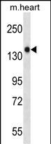
Mouse Gucy2f Antibody (Center)
Affinity Purified Rabbit Polyclonal Antibody (Pab)
- SPECIFICATION
- CITATIONS
- PROTOCOLS
- BACKGROUND

Application
| WB, E |
|---|---|
| Primary Accession | Q5SDA5 |
| Other Accession | NP_001007577.1 |
| Reactivity | Mouse |
| Host | Rabbit |
| Clonality | Polyclonal |
| Isotype | Rabbit IgG |
| Calculated MW | 124425 Da |
| Antigen Region | 317-344 aa |
| Gene ID | 245650 |
|---|---|
| Other Names | Retinal guanylyl cyclase 2, Gucy2f |
| Target/Specificity | This Mouse Gucy2f antibody is generated from rabbits immunized with a KLH conjugated synthetic peptide between 317-344 amino acids from the Central region of mouse Gucy2f. |
| Dilution | WB~~1:1000 E~~Use at an assay dependent concentration. |
| Format | Purified polyclonal antibody supplied in PBS with 0.09% (W/V) sodium azide. This antibody is purified through a protein A column, followed by peptide affinity purification. |
| Storage | Maintain refrigerated at 2-8°C for up to 2 weeks. For long term storage store at -20°C in small aliquots to prevent freeze-thaw cycles. |
| Precautions | Mouse Gucy2f Antibody (Center) is for research use only and not for use in diagnostic or therapeutic procedures. |
| Name | Gucy2f {ECO:0000312|MGI:MGI:105119} |
|---|---|
| Function | Responsible for the synthesis of cyclic GMP (cGMP) in rods and cones of photoreceptors (By similarity). Plays an essential role in phototransduction, by mediating cGMP replenishment. May also participate in the trafficking of membrane-asociated proteins to the photoreceptor outer segment membrane (PubMed:17255100). |
| Cellular Location | Membrane {ECO:0000250|UniProtKB:P51842}; Single- pass type I membrane protein. Photoreceptor outer segment membrane; Single-pass type I membrane protein |
| Tissue Location | Retina.. |

Thousands of laboratories across the world have published research that depended on the performance of antibodies from Abcepta to advance their research. Check out links to articles that cite our products in major peer-reviewed journals, organized by research category.
info@abcepta.com, and receive a free "I Love Antibodies" mug.

Provided below are standard protocols that you may find useful for product applications.
Background
Probably plays a specific functional role in the rods and/or cones of photoreceptors. It may be the enzyme involved in the resynthesis of cGMP required for recovery of the dark state after phototransduction (By similarity).
References
Schmidt, H., et al. J. Cell Biol. 179(2):331-340(2007)
Baehr, W., et al. J. Biol. Chem. 282(12):8837-8847(2007)
Corbo, J.C., et al. PLoS Genet. 1 (2), E11 (2005) :
Shearstone, J.R., et al. Genomics 85(3):309-321(2005)
Yamazaki, K., et al. Genomics 51(2):303-305(1998)
If you have used an Abcepta product and would like to share how it has performed, please click on the "Submit Review" button and provide the requested information. Our staff will examine and post your review and contact you if needed.
If you have any additional inquiries please email technical services at tech@abcepta.com.
Ordering Information
Other Products
Shipping Information
