


 Foundational characteristics of cancer include proliferation, angiogenesis, migration, evasion of apoptosis, and cellular immortality. Find key markers for these cellular processes and antibodies to detect them.
 The SUMOplot™ Analysis Program predicts and scores sumoylation sites in your protein. SUMOylation is a post-translational modification involved in various cellular processes, such as nuclear-cytosolic transport, transcriptional regulation, apoptosis, protein stability, response to stress, and progression through the cell cycle.
 The Autophagy Receptor Motif Plotter predicts and scores autophagy receptor binding sites in your protein. Identifying proteins connected to this pathway is critical to understanding the role of autophagy in physiological as well as pathological processes such as development, differentiation, neurodegenerative diseases, stress, infection, and cancer.
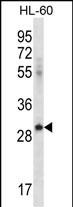
ICOSLG Antibody (C-term)
Affinity Purified Rabbit Polyclonal Antibody (Pab)
- SPECIFICATION
- CITATIONS
- PROTOCOLS
- BACKGROUND

Application
| WB, E |
|---|---|
| Primary Accession | O75144 |
| Other Accession | NP_056074.1 |
| Reactivity | Human |
| Host | Rabbit |
| Clonality | Polyclonal |
| Isotype | Rabbit IgG |
| Calculated MW | 33349 Da |
| Antigen Region | 217-246 aa |
| Gene ID | 23308 |
|---|---|
| Other Names | ICOS ligand, B7 homolog 2, B7-H2, B7-like protein Gl50, B7-related protein 1, B7RP-1, CD275, ICOSLG, B7H2, B7RP1, ICOSL, KIAA0653 |
| Target/Specificity | This ICOSLG antibody is generated from rabbits immunized with a KLH conjugated synthetic peptide between 217-246 amino acids from the C-terminal region of human ICOSLG. |
| Dilution | WB~~1:1000 E~~Use at an assay dependent concentration. |
| Format | Purified polyclonal antibody supplied in PBS with 0.09% (W/V) sodium azide. This antibody is purified through a protein A column, followed by peptide affinity purification. |
| Storage | Maintain refrigerated at 2-8°C for up to 2 weeks. For long term storage store at -20°C in small aliquots to prevent freeze-thaw cycles. |
| Precautions | ICOSLG Antibody (C-term) is for research use only and not for use in diagnostic or therapeutic procedures. |
| Name | ICOSLG |
|---|---|
| Function | Ligand for the T-cell-specific cell surface receptor ICOS. Acts as a costimulatory signal for T-cell proliferation and cytokine secretion (PubMed:11007762, PubMed:11023515, PubMed:30498080). Also induces B-cell proliferation and differentiation into plasma cells. Could play an important role in mediating local tissue responses to inflammatory conditions, as well as in modulating the secondary immune response by co-stimulating memory T-cell function (By similarity). In endothelial cells, required for proper neutrophil transmigration in response to chemoattractants, such as CXCL8/IL8 or N-formyl-methionyl peptides (fMLP) (PubMed:30498080). |
| Cellular Location | Cell membrane; Single-pass type I membrane protein |
| Tissue Location | Expressed on peripheral blood B-cells and monocytes, as well as on monocyte-derived dendritic cells (at protein level). [Isoform 2]: Detected only in lymph nodes, leukocytes and spleen. Expressed on activated monocytes and dendritic cells. |

Thousands of laboratories across the world have published research that depended on the performance of antibodies from Abcepta to advance their research. Check out links to articles that cite our products in major peer-reviewed journals, organized by research category.
info@abcepta.com, and receive a free "I Love Antibodies" mug.

Provided below are standard protocols that you may find useful for product applications.
Background
Ligand for the T-cell-specific cell surface receptor ICOS. Acts as a costimulatory signal for T-cell proliferation and cytokine secretion; induces also B-cell proliferation and differentiation into plasma cells. Could play an important role in mediating local tissue responses to inflammatory conditions, as well as in modulating the secondary immune response by co-stimulating memory T-cell function (By similarity).
References
Shimada, M., et al. Hum. Genet. 128(4):433-441(2010)
Bailey, S.D., et al. Diabetes Care 33(10):2250-2253(2010)
Wang, K., et al. Hum. Mol. Genet. 19(10):2059-2067(2010)
Pan, F., et al. Immunogenetics 62(4):237-251(2010)
Dubois, P.C., et al. Nat. Genet. 42(4):295-302(2010)
If you have used an Abcepta product and would like to share how it has performed, please click on the "Submit Review" button and provide the requested information. Our staff will examine and post your review and contact you if needed.
If you have any additional inquiries please email technical services at tech@abcepta.com.
Ordering Information
Other Products
Shipping Information
