


 Foundational characteristics of cancer include proliferation, angiogenesis, migration, evasion of apoptosis, and cellular immortality. Find key markers for these cellular processes and antibodies to detect them.
 The SUMOplot™ Analysis Program predicts and scores sumoylation sites in your protein. SUMOylation is a post-translational modification involved in various cellular processes, such as nuclear-cytosolic transport, transcriptional regulation, apoptosis, protein stability, response to stress, and progression through the cell cycle.
 The Autophagy Receptor Motif Plotter predicts and scores autophagy receptor binding sites in your protein. Identifying proteins connected to this pathway is critical to understanding the role of autophagy in physiological as well as pathological processes such as development, differentiation, neurodegenerative diseases, stress, infection, and cancer.
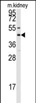
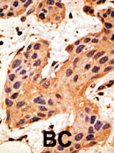
Connexin 50 Antibody (C-term)
Purified Rabbit Polyclonal Antibody (Pab)
- SPECIFICATION
- CITATIONS
- PROTOCOLS
- BACKGROUND

Application
| IHC-P, WB, E |
|---|---|
| Primary Accession | P48165 |
| Other Accession | P36381, P55917 |
| Reactivity | Human, Mouse |
| Predicted | Chicken, Sheep |
| Host | Rabbit |
| Clonality | Polyclonal |
| Isotype | Rabbit IgG |
| Calculated MW | 48229 Da |
| Antigen Region | 402-433 aa |
| Gene ID | 2703 |
|---|---|
| Other Names | Gap junction alpha-8 protein, Connexin-50, Cx50, Lens fiber protein MP70, GJA8 |
| Target/Specificity | This Connexin 50 antibody is generated from rabbits immunized with a KLH conjugated synthetic peptide between 402-433 amino acids from the C-terminal region of human Connexin 50. |
| Dilution | IHC-P~~1:50~100 WB~~1:1000 E~~Use at an assay dependent concentration. |
| Format | Purified polyclonal antibody supplied in PBS with 0.09% (W/V) sodium azide. This antibody is prepared by Saturated Ammonium Sulfate (SAS) precipitation followed by dialysis against PBS. |
| Storage | Maintain refrigerated at 2-8°C for up to 2 weeks. For long term storage store at -20°C in small aliquots to prevent freeze-thaw cycles. |
| Precautions | Connexin 50 Antibody (C-term) is for research use only and not for use in diagnostic or therapeutic procedures. |
| Name | GJA8 |
|---|---|
| Function | Structural component of eye lens gap junctions (PubMed:18006672, PubMed:19756179). Gap junctions are dodecameric channels that connect the cytoplasm of adjoining cells. They are formed by the docking of two hexameric hemichannels, one from each cell membrane (By similarity). Small molecules and ions diffuse from one cell to a neighboring cell via the central pore (PubMed:18006672, PubMed:19756179). |
| Cellular Location | Cell membrane; Multi-pass membrane protein {ECO:0000250|UniProtKB:P55917}. Cell junction, gap junction |
| Tissue Location | Eye lens.. |

Thousands of laboratories across the world have published research that depended on the performance of antibodies from Abcepta to advance their research. Check out links to articles that cite our products in major peer-reviewed journals, organized by research category.
info@abcepta.com, and receive a free "I Love Antibodies" mug.

Provided below are standard protocols that you may find useful for product applications.
Background
GJA8 is a an integral membrane protein that belongs to the connexin family, alpha-type (group II) subfamily. One gap junction consists of a cluster of closely packed pairs of transmembrane channels, the connexons, through which materials of low MW diffuse from one cell to a neighboring cell. A connexon is composed of a hexamer of connexins. This particular connexin only forms junctional channels. GJA8 is expressed in the eye lens, and defects in GJA8 are the cause of zonular pulverulent cataract type 1 (CZP1), a form of autosomal dominant congenital cataract.
References
Shiels, A., et al., Am. J. Hum. Genet. 62(3):526-532 (1998). Church, R.L., et al., Curr. Eye Res. 14(10):979-981 (1995). Church, R.L., et al., Curr. Eye Res. 14(3):215-221 (1995).
If you have used an Abcepta product and would like to share how it has performed, please click on the "Submit Review" button and provide the requested information. Our staff will examine and post your review and contact you if needed.
If you have any additional inquiries please email technical services at tech@abcepta.com.
Ordering Information
Other Products
Shipping Information
