


 Foundational characteristics of cancer include proliferation, angiogenesis, migration, evasion of apoptosis, and cellular immortality. Find key markers for these cellular processes and antibodies to detect them.
 The SUMOplot™ Analysis Program predicts and scores sumoylation sites in your protein. SUMOylation is a post-translational modification involved in various cellular processes, such as nuclear-cytosolic transport, transcriptional regulation, apoptosis, protein stability, response to stress, and progression through the cell cycle.
 The Autophagy Receptor Motif Plotter predicts and scores autophagy receptor binding sites in your protein. Identifying proteins connected to this pathway is critical to understanding the role of autophagy in physiological as well as pathological processes such as development, differentiation, neurodegenerative diseases, stress, infection, and cancer.
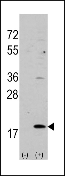
VILIP3 Antibody (N-term)
Purified Rabbit Polyclonal Antibody (Pab)


- SPECIFICATION
- CITATIONS
- PROTOCOLS
- BACKGROUND

Application
| WB, IHC-P, E |
|---|---|
| Primary Accession | P37235 |
| Reactivity | Human, Mouse |
| Host | Rabbit |
| Clonality | Polyclonal |
| Isotype | Rabbit IgG |
| Calculated MW | 22313 Da |
| Antigen Region | 2-32 aa |
| Gene ID | 3241 |
|---|---|
| Other Names | Hippocalcin-like protein 1, Calcium-binding protein BDR-1, HLP2, Visinin-like protein 3, VILIP-3, HPCAL1, BDR1 |
| Target/Specificity | This VILIP3 antibody is generated from rabbits immunized with a KLH conjugated synthetic peptide between 2-32 amino acids from the N-terminal region of human VILIP3. |
| Dilution | WB~~1:1000 IHC-P~~1:50~100 E~~Use at an assay dependent concentration. |
| Format | Purified polyclonal antibody supplied in PBS with 0.09% (W/V) sodium azide. This antibody is prepared by Saturated Ammonium Sulfate (SAS) precipitation followed by dialysis against PBS. |
| Storage | Maintain refrigerated at 2-8°C for up to 2 weeks. For long term storage store at -20°C in small aliquots to prevent freeze-thaw cycles. |
| Precautions | VILIP3 Antibody (N-term) is for research use only and not for use in diagnostic or therapeutic procedures. |
| Name | HPCAL1 |
|---|---|
| Synonyms | BDR1 |
| Function | May be involved in the calcium-dependent regulation of rhodopsin phosphorylation. |
| Cellular Location | Membrane; Lipid-anchor |

Thousands of laboratories across the world have published research that depended on the performance of antibodies from Abcepta to advance their research. Check out links to articles that cite our products in major peer-reviewed journals, organized by research category.
info@abcepta.com, and receive a free "I Love Antibodies" mug.

Provided below are standard protocols that you may find useful for product applications.
Background
VILIP3 is a member of neuron-specific calcium-binding proteins family found in the retina and brain. It is highly similar to human hippocalcin protein and nearly identical to the rat and mouse hippocalcin like-1 proteins. It may be involved in the calcium-dependent regulation of rhodopsin phosphorylation and may be of relevance for neuronal signalling in the central nervous system. There are two alternatively spliced transcript variants of this gene, with multiple polyadenylation sites. Transcript variant 1 utilizes a different exon and also lacks one exon in the 5' UTR, as compared to variant 2; thus, the encoded protein is the same.
References
Braunewell, K., et al., Dement Geriatr Cogn Disord 12(2):110-116 (2001). Bernstein, H.G., et al., J Neurocytol 28(8):655-662 (1999). Kobayashi, M., et al., Biochim. Biophys. Acta 1222(3):515-518 (1994). Hidaka, H., et al., Neurosci. Res. 16(2):73-77 (1993). Ivings, L., et al., Biochem. J. 363 (Pt 3), 599-608 (2002).
If you have used an Abcepta product and would like to share how it has performed, please click on the "Submit Review" button and provide the requested information. Our staff will examine and post your review and contact you if needed.
If you have any additional inquiries please email technical services at tech@abcepta.com.
Ordering Information
Other Products
Shipping Information
