


 Foundational characteristics of cancer include proliferation, angiogenesis, migration, evasion of apoptosis, and cellular immortality. Find key markers for these cellular processes and antibodies to detect them.
 The SUMOplot™ Analysis Program predicts and scores sumoylation sites in your protein. SUMOylation is a post-translational modification involved in various cellular processes, such as nuclear-cytosolic transport, transcriptional regulation, apoptosis, protein stability, response to stress, and progression through the cell cycle.
 The Autophagy Receptor Motif Plotter predicts and scores autophagy receptor binding sites in your protein. Identifying proteins connected to this pathway is critical to understanding the role of autophagy in physiological as well as pathological processes such as development, differentiation, neurodegenerative diseases, stress, infection, and cancer.
FAU Antibody
Affinity Purified Rabbit Polyclonal Antibody (Pab)
- SPECIFICATION
- CITATIONS
- PROTOCOLS
- BACKGROUND

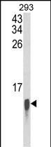
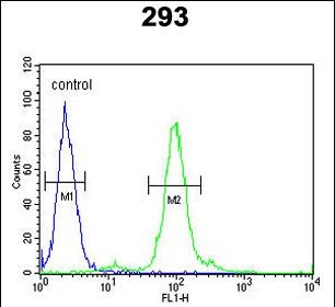
Application
| WB, FC, E |
|---|---|
| Primary Accession | P35544 |
| Reactivity | Human |
| Host | Rabbit |
| Clonality | Polyclonal |
| Isotype | Rabbit IgG |
| Other Names | Ubiquitin-like protein FUBI, FAU |
|---|---|
| Target/Specificity | This FAU antibody is generated from rabbits immunized with human FAU recombinant protein. |
| Dilution | WB~~1:1000 FC~~1:10~50 E~~Use at an assay dependent concentration. |
| Format | Purified polyclonal antibody supplied in PBS with 0.09% (W/V) sodium azide. This antibody is purified through a protein A column, followed by peptide affinity purification. |
| Storage | Maintain refrigerated at 2-8°C for up to 2 weeks. For long term storage store at -20°C in small aliquots to prevent freeze-thaw cycles. |
| Precautions | FAU Antibody is for research use only and not for use in diagnostic or therapeutic procedures. |

Thousands of laboratories across the world have published research that depended on the performance of antibodies from Abcepta to advance their research. Check out links to articles that cite our products in major peer-reviewed journals, organized by research category.
info@abcepta.com, and receive a free "I Love Antibodies" mug.

Provided below are standard protocols that you may find useful for product applications.
Background
FUBI is the cellular homolog of the fox sequence in the Finkel-Biskis-Reilly murine sarcoma virus (FBR-MuSV). It is a fusion protein consisting of the ubiquitin-like protein fubi at the N terminus and ribosomal protein S30 at the C terminus. It has been proposed that the fusion protein is post-translationally processed to generate free fubi and free ribosomal protein S30. Fubi is a member of the ubiquitin family, and ribosomal protein S30 belongs to the S30E family of ribosomal proteins. Whereas the function of fubi is currently unknown, ribosomal protein S30 is a component of the 40S subunit of the cytoplasmic ribosome.
References
Rossman, T.G., et al., Oncogene 22(12):1817-1821 (2003).
Kenmochi, N., et al., Genome Res. 8(5):509-523 (1998).
Vladimirov, S.N., et al., Eur. J. Biochem. 239(1):144-149 (1996).
Kas, K., et al., Genomics 17(2):387-392 (1993).
Michiels, L., et al., Oncogene 8(9):2537-2546 (1993).
If you have used an Abcepta product and would like to share how it has performed, please click on the "Submit Review" button and provide the requested information. Our staff will examine and post your review and contact you if needed.
If you have any additional inquiries please email technical services at tech@abcepta.com.
Ordering Information
Shipping Information
