


 Foundational characteristics of cancer include proliferation, angiogenesis, migration, evasion of apoptosis, and cellular immortality. Find key markers for these cellular processes and antibodies to detect them.
 The SUMOplot™ Analysis Program predicts and scores sumoylation sites in your protein. SUMOylation is a post-translational modification involved in various cellular processes, such as nuclear-cytosolic transport, transcriptional regulation, apoptosis, protein stability, response to stress, and progression through the cell cycle.
 The Autophagy Receptor Motif Plotter predicts and scores autophagy receptor binding sites in your protein. Identifying proteins connected to this pathway is critical to understanding the role of autophagy in physiological as well as pathological processes such as development, differentiation, neurodegenerative diseases, stress, infection, and cancer.
RBBP7 Antibody (Center)
Affinity Purified Rabbit Polyclonal Antibody (Pab)
- SPECIFICATION
- CITATIONS
- PROTOCOLS
- BACKGROUND

Application
| WB, E |
|---|---|
| Primary Accession | Q16576 |
| Other Accession | Q71UF4, Q60973, Q4R304, Q3SWX8, NP_002884.1 |
| Reactivity | Human |
| Predicted | Bovine, Monkey, Mouse, Rat |
| Host | Rabbit |
| Clonality | Polyclonal |
| Isotype | Rabbit IgG |
| Calculated MW | 47820 Da |
| Antigen Region | 191-219 aa |
| Gene ID | 5931 |
|---|---|
| Other Names | Histone-binding protein RBBP7, Histone acetyltransferase type B subunit 2, Nucleosome-remodeling factor subunit RBAP46, Retinoblastoma-binding protein 7, RBBP-7, Retinoblastoma-binding protein p46, RBBP7, RBAP46 |
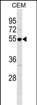
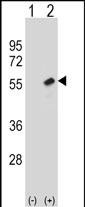
| Target/Specificity | This RBBP7 antibody is generated from rabbits immunized with a KLH conjugated synthetic peptide between 191-219 amino acids from the Central region of human RBBP7. |
| Dilution | WB~~1:1000 E~~Use at an assay dependent concentration. |
| Format | Purified polyclonal antibody supplied in PBS with 0.09% (W/V) sodium azide. This antibody is purified through a protein A column, followed by peptide affinity purification. |
| Storage | Maintain refrigerated at 2-8°C for up to 2 weeks. For long term storage store at -20°C in small aliquots to prevent freeze-thaw cycles. |
| Precautions | RBBP7 Antibody (Center) is for research use only and not for use in diagnostic or therapeutic procedures. |
| Name | RBBP7 |
|---|---|
| Synonyms | RBAP46 |
| Function | Core histone-binding subunit that may target chromatin remodeling factors, histone acetyltransferases and histone deacetylases to their histone substrates in a manner that is regulated by nucleosomal DNA. Component of several complexes which regulate chromatin metabolism. These include the type B histone acetyltransferase (HAT) complex, which is required for chromatin assembly following DNA replication; the core histone deacetylase (HDAC) complex, which promotes histone deacetylation and consequent transcriptional repression; the nucleosome remodeling and histone deacetylase complex (the NuRD complex), which promotes transcriptional repression by histone deacetylation and nucleosome remodeling; and the PRC2/EED-EZH2 complex, which promotes repression of homeotic genes during development; and the NURF (nucleosome remodeling factor) complex. |
| Cellular Location | Nucleus |

Thousands of laboratories across the world have published research that depended on the performance of antibodies from Abcepta to advance their research. Check out links to articles that cite our products in major peer-reviewed journals, organized by research category.
info@abcepta.com, and receive a free "I Love Antibodies" mug.

Provided below are standard protocols that you may find useful for product applications.
Background
This protein is a ubiquitously expressed nuclear protein and belongs to a highly conserved subfamily of WD-repeat proteins. It is found among several proteins that binds directly to retinoblastoma protein, which regulates cell proliferation. The encoded protein is found in many histone deacetylase complexes, including mSin3 co-repressor complex. It is also present in protein complexes involved in chromatin assembly. This protein can interact with BRCA1 tumor-suppressor gene and may have a role in the regulation of cell proliferation and differentiation. Two transcript variants encoding different isoforms have been found for this gene.
References
Bailey, S.D., et al. Diabetes Care 33(10):2250-2253(2010)
Talmud, P.J., et al. Am. J. Hum. Genet. 85(5):628-642(2009)
Saade, E., et al. Proteomics 9(21):4934-4943(2009)
Wang, C.L., et al. J. Proteome Res. 8(10):4428-4440(2009)
Li, R., et al. EMBO J. 28(18):2763-2776(2009)
If you have used an Abcepta product and would like to share how it has performed, please click on the "Submit Review" button and provide the requested information. Our staff will examine and post your review and contact you if needed.
If you have any additional inquiries please email technical services at tech@abcepta.com.
Ordering Information
Other Products
Shipping Information
