


 Foundational characteristics of cancer include proliferation, angiogenesis, migration, evasion of apoptosis, and cellular immortality. Find key markers for these cellular processes and antibodies to detect them.
 The SUMOplot™ Analysis Program predicts and scores sumoylation sites in your protein. SUMOylation is a post-translational modification involved in various cellular processes, such as nuclear-cytosolic transport, transcriptional regulation, apoptosis, protein stability, response to stress, and progression through the cell cycle.
 The Autophagy Receptor Motif Plotter predicts and scores autophagy receptor binding sites in your protein. Identifying proteins connected to this pathway is critical to understanding the role of autophagy in physiological as well as pathological processes such as development, differentiation, neurodegenerative diseases, stress, infection, and cancer.
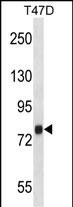
ZC3H14 Antibody (Center)
Affinity Purified Rabbit Polyclonal Antibody (Pab)
- SPECIFICATION
- CITATIONS
- PROTOCOLS
- BACKGROUND

Application
| WB, E |
|---|---|
| Primary Accession | Q6PJT7 |
| Other Accession | Q4R6F6, NP_997545.2 |
| Reactivity | Human |
| Predicted | Monkey |
| Host | Rabbit |
| Clonality | Polyclonal |
| Isotype | Rabbit IgG |
| Calculated MW | 82876 Da |
| Antigen Region | 401-429 aa |
| Gene ID | 79882 |
|---|---|
| Other Names | Zinc finger CCCH domain-containing protein 14, Mammalian suppressor of tau pathology-2, MSUT-2, Renal carcinoma antigen NY-REN-37, ZC3H14 |
| Target/Specificity | This ZC3H14 antibody is generated from rabbits immunized with a KLH conjugated synthetic peptide between 401-429 amino acids from the Central region of human ZC3H14. |
| Dilution | WB~~1:1000 E~~Use at an assay dependent concentration. |
| Format | Purified polyclonal antibody supplied in PBS with 0.09% (W/V) sodium azide. This antibody is purified through a protein A column, followed by peptide affinity purification. |
| Storage | Maintain refrigerated at 2-8°C for up to 2 weeks. For long term storage store at -20°C in small aliquots to prevent freeze-thaw cycles. |
| Precautions | ZC3H14 Antibody (Center) is for research use only and not for use in diagnostic or therapeutic procedures. |
| Name | ZC3H14 {ECO:0000303|PubMed:19303045, ECO:0000312|HGNC:HGNC:20509} |
|---|---|
| Function | RNA-binding protein involved in the biogenesis of circular RNAs (circRNAs), which are produced by back-splicing circularization of pre-mRNAs (PubMed:39461343). Acts by binding to both exon-intron boundary and 3'-UTR of pre-mRNAs to promote circRNA biogenesis through dimerization and the association with the spliceosome (PubMed:39461343). Required for spermatogenesis via involvement in circRNA biogenesis (PubMed:39461343). Regulates the pre-mRNA processing of ATP5MC1; preventing its degradation (PubMed:27563065). Also binds the poly(A) tail of mRNAs; controlling poly(A) length in neuronal cells (PubMed:17630287, PubMed:24671764). |
| Cellular Location | Nucleus speckle Note=Colocalizes with poly(A) RNA in nuclear speckles {ECO:0000250|UniProtKB:Q7TMD5} [Isoform 3]: Nucleus speckle |
| Tissue Location | [Isoform 1]: Expressed in fetal and adult brain (PubMed:21734151). Expressed in fetal and adult temporal lobe (PubMed:21734151). |

Thousands of laboratories across the world have published research that depended on the performance of antibodies from Abcepta to advance their research. Check out links to articles that cite our products in major peer-reviewed journals, organized by research category.
info@abcepta.com, and receive a free "I Love Antibodies" mug.

Provided below are standard protocols that you may find useful for product applications.
Background
ZC3H14 belongs to a family of poly(A)-binding proteins that influence gene expression by regulating mRNA stability, nuclear export, and translation (Kelly et al., 2007 [PubMed 17630287]).
References
Wheeler, J.M., et al. Biochem. Soc. Trans. 38(4):973-976(2010)
Leung, S.W., et al. Gene 439 (1-2), 71-78 (2009) :
Kelly, S.M., et al. Proc. Natl. Acad. Sci. U.S.A. 104(30):12306-12311(2007)
Scanlan, M.J., et al. Int. J. Cancer 83(4):456-464(1999)
If you have used an Abcepta product and would like to share how it has performed, please click on the "Submit Review" button and provide the requested information. Our staff will examine and post your review and contact you if needed.
If you have any additional inquiries please email technical services at tech@abcepta.com.
Ordering Information
Other Products
Shipping Information
