


 Foundational characteristics of cancer include proliferation, angiogenesis, migration, evasion of apoptosis, and cellular immortality. Find key markers for these cellular processes and antibodies to detect them.
 The SUMOplot™ Analysis Program predicts and scores sumoylation sites in your protein. SUMOylation is a post-translational modification involved in various cellular processes, such as nuclear-cytosolic transport, transcriptional regulation, apoptosis, protein stability, response to stress, and progression through the cell cycle.
 The Autophagy Receptor Motif Plotter predicts and scores autophagy receptor binding sites in your protein. Identifying proteins connected to this pathway is critical to understanding the role of autophagy in physiological as well as pathological processes such as development, differentiation, neurodegenerative diseases, stress, infection, and cancer.
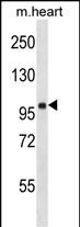
FHOD1 Antibody (C-term)
Affinity Purified Rabbit Polyclonal Antibody (Pab)
- SPECIFICATION
- CITATIONS
- PROTOCOLS
- BACKGROUND

Application
| WB, E |
|---|---|
| Primary Accession | Q9Y613 |
| Other Accession | NP_037373.2 |
| Reactivity | Human, Mouse |
| Host | Rabbit |
| Clonality | Polyclonal |
| Isotype | Rabbit IgG |
| Calculated MW | 126551 Da |
| Antigen Region | 985-1013 aa |
| Gene ID | 29109 |
|---|---|
| Other Names | FH1/FH2 domain-containing protein 1, Formin homolog overexpressed in spleen 1, FHOS, Formin homology 2 domain-containing protein 1, FHOD1, FHOS, FHOS1 |
| Target/Specificity | This FHOD1 antibody is generated from rabbits immunized with a KLH conjugated synthetic peptide between 985-1013 amino acids from the C-terminal region of human FHOD1. |
| Dilution | WB~~1:1000 E~~Use at an assay dependent concentration. |
| Format | Purified polyclonal antibody supplied in PBS with 0.09% (W/V) sodium azide. This antibody is purified through a protein A column, followed by peptide affinity purification. |
| Storage | Maintain refrigerated at 2-8°C for up to 2 weeks. For long term storage store at -20°C in small aliquots to prevent freeze-thaw cycles. |
| Precautions | FHOD1 Antibody (C-term) is for research use only and not for use in diagnostic or therapeutic procedures. |
| Name | FHOD1 |
|---|---|
| Synonyms | FHOS, FHOS1 |
| Function | Required for the assembly of F-actin structures, such as stress fibers. Depends on the Rho-ROCK cascade for its activity. Contributes to the coordination of microtubules with actin fibers and plays a role in cell elongation. Acts synergistically with ROCK1 to promote SRC-dependent non-apoptotic plasma membrane blebbing. |
| Cellular Location | Cytoplasm. Cytoplasm, cytoskeleton. Cell projection, bleb. Note=Predominantly cytoplasmic |
| Tissue Location | Ubiquitous. Highly expressed in spleen. |

Thousands of laboratories across the world have published research that depended on the performance of antibodies from Abcepta to advance their research. Check out links to articles that cite our products in major peer-reviewed journals, organized by research category.
info@abcepta.com, and receive a free "I Love Antibodies" mug.

Provided below are standard protocols that you may find useful for product applications.
Background
This gene encodes a protein which is a member of the formin/diaphanous family of proteins. The gene is ubiquitously expressed but is found in abundance in the spleen. The encoded protein has sequence homology to diaphanous and formin proteins within the Formin Homology (FH)1 and FH2 domains. It also contains a coiled-coil domain, a collagen-like domain, two nuclear localization signals, and several potential PKC and PKA phosphorylation sites. It is a predominantly cytoplasmic protein and is expressed in a variety of human cell lines. [provided by RefSeq].
References
Hannemann, S., et al. J. Biol. Chem. 283(41):27891-27903(2008)
Schulte, A., et al. Structure 16(9):1313-1323(2008)
Takeya, R., et al. EMBO J. 27(4):618-628(2008)
Schulte, A., et al. Acta Crystallogr. Sect. F Struct. Biol. Cryst. Commun. 63 (PT 10), 878-881 (2007) :
Sugiyama, N., et al. Mol. Cell Proteomics 6(6):1103-1109(2007)
If you have used an Abcepta product and would like to share how it has performed, please click on the "Submit Review" button and provide the requested information. Our staff will examine and post your review and contact you if needed.
If you have any additional inquiries please email technical services at tech@abcepta.com.
Ordering Information
Other Products
Shipping Information
