


 Foundational characteristics of cancer include proliferation, angiogenesis, migration, evasion of apoptosis, and cellular immortality. Find key markers for these cellular processes and antibodies to detect them.
 The SUMOplot™ Analysis Program predicts and scores sumoylation sites in your protein. SUMOylation is a post-translational modification involved in various cellular processes, such as nuclear-cytosolic transport, transcriptional regulation, apoptosis, protein stability, response to stress, and progression through the cell cycle.
 The Autophagy Receptor Motif Plotter predicts and scores autophagy receptor binding sites in your protein. Identifying proteins connected to this pathway is critical to understanding the role of autophagy in physiological as well as pathological processes such as development, differentiation, neurodegenerative diseases, stress, infection, and cancer.
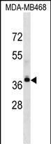
LPAR4 Antibody (Center)
Affinity Purified Rabbit Polyclonal Antibody (Pab)
- SPECIFICATION
- CITATIONS
- PROTOCOLS
- BACKGROUND

Application
| WB, E |
|---|---|
| Primary Accession | Q99677 |
| Other Accession | Q8BLG2, NP_005287.1 |
| Reactivity | Human |
| Predicted | Mouse |
| Host | Rabbit |
| Clonality | Polyclonal |
| Isotype | Rabbit IgG |
| Calculated MW | 41895 Da |
| Antigen Region | 127-156 aa |
| Gene ID | 2846 |
|---|---|
| Other Names | Lysophosphatidic acid receptor 4, LPA receptor 4, LPA-4, G-protein coupled receptor 23, P2Y purinoceptor 9, P2Y9, P2Y5-like receptor, Purinergic receptor 9, LPAR4, GPR23, LPA4, P2RY9 |
| Target/Specificity | This LPAR4 antibody is generated from rabbits immunized with a KLH conjugated synthetic peptide between 127-156 amino acids from the Central region of human LPAR4. |
| Dilution | WB~~1:1000 E~~Use at an assay dependent concentration. |
| Format | Purified polyclonal antibody supplied in PBS with 0.09% (W/V) sodium azide. This antibody is purified through a protein A column, followed by peptide affinity purification. |
| Storage | Maintain refrigerated at 2-8°C for up to 2 weeks. For long term storage store at -20°C in small aliquots to prevent freeze-thaw cycles. |
| Precautions | LPAR4 Antibody (Center) is for research use only and not for use in diagnostic or therapeutic procedures. |
| Name | LPAR4 |
|---|---|
| Synonyms | GPR23, LPA4, P2RY9 |
| Function | Receptor for lysophosphatidic acid (LPA), a mediator of diverse cellular activities. Transduces a signal by increasing the intracellular calcium ions and by stimulating adenylyl cyclase activity. The rank order of potency for agonists of this receptor is 1- oleoyl- > 1-stearoyl- > 1-palmitoyl- > 1-myristoyl- > 1-alkyl- > 1- alkenyl-LPA. |
| Cellular Location | Cell membrane; Multi-pass membrane protein. |
| Tissue Location | High expression in ovary. Not detected in the brain regions thalamus, putamen, caudate, frontal cortex, pons, hypothalamus and hippocampus. |

Thousands of laboratories across the world have published research that depended on the performance of antibodies from Abcepta to advance their research. Check out links to articles that cite our products in major peer-reviewed journals, organized by research category.
info@abcepta.com, and receive a free "I Love Antibodies" mug.

Provided below are standard protocols that you may find useful for product applications.
Background
This gene encodes a member of the lysophosphatidic acid receptor family. It may also be related to the P2Y receptors, a family of receptors that bind purine and pyrimidine nucleotides and are coupled to G proteins. The encoded protein may play a role in monocytic differentiation.
References
Liu, Y.B., et al. J. Cell. Biochem. 109(4):794-800(2010)
Kumar, S.A., et al. Leuk. Lymphoma 50(12):2038-2048(2009)
Luttrell, L.M. Mol. Biotechnol. 39(3):239-264(2008)
Dowal, L., et al. J. Biol. Chem. 281(33):23999-24014(2006)
Noguchi, K., et al. J. Biol. Chem. 278(28):25600-25606(2003)
If you have used an Abcepta product and would like to share how it has performed, please click on the "Submit Review" button and provide the requested information. Our staff will examine and post your review and contact you if needed.
If you have any additional inquiries please email technical services at tech@abcepta.com.
Ordering Information
Other Products
Shipping Information
