


 Foundational characteristics of cancer include proliferation, angiogenesis, migration, evasion of apoptosis, and cellular immortality. Find key markers for these cellular processes and antibodies to detect them.
 The SUMOplot™ Analysis Program predicts and scores sumoylation sites in your protein. SUMOylation is a post-translational modification involved in various cellular processes, such as nuclear-cytosolic transport, transcriptional regulation, apoptosis, protein stability, response to stress, and progression through the cell cycle.
 The Autophagy Receptor Motif Plotter predicts and scores autophagy receptor binding sites in your protein. Identifying proteins connected to this pathway is critical to understanding the role of autophagy in physiological as well as pathological processes such as development, differentiation, neurodegenerative diseases, stress, infection, and cancer.
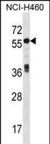
ICA1 Antibody (N-term)
Affinity Purified Rabbit Polyclonal Antibody (Pab)
- SPECIFICATION
- CITATIONS
- PROTOCOLS
- BACKGROUND

Application
| WB, E |
|---|---|
| Primary Accession | Q05084 |
| Other Accession | Q63054, P97411, NP_071682.1, NP_001129492.1 |
| Reactivity | Human |
| Predicted | Mouse, Rat |
| Host | Rabbit |
| Clonality | Polyclonal |
| Isotype | Rabbit IgG |
| Calculated MW | 54645 Da |
| Antigen Region | 84-111 aa |
| Gene ID | 3382 |
|---|---|
| Other Names | Islet cell autoantigen 1, 69 kDa islet cell autoantigen, ICA69, Islet cell autoantigen p69, ICAp69, p69, ICA1 |
| Target/Specificity | This ICA1 antibody is generated from rabbits immunized with a KLH conjugated synthetic peptide between 84-111 amino acids from the N-terminal region of human ICA1. |
| Dilution | WB~~1:1000 E~~Use at an assay dependent concentration. |
| Format | Purified polyclonal antibody supplied in PBS with 0.09% (W/V) sodium azide. This antibody is purified through a protein A column, followed by peptide affinity purification. |
| Storage | Maintain refrigerated at 2-8°C for up to 2 weeks. For long term storage store at -20°C in small aliquots to prevent freeze-thaw cycles. |
| Precautions | ICA1 Antibody (N-term) is for research use only and not for use in diagnostic or therapeutic procedures. |
| Name | ICA1 |
|---|---|
| Function | May play a role in neurotransmitter secretion. |
| Cellular Location | Cytoplasm, cytosol. Golgi apparatus membrane; Peripheral membrane protein. Cytoplasmic vesicle, secretory vesicle membrane; Peripheral membrane protein. Cytoplasmic vesicle, secretory vesicle, synaptic vesicle membrane; Peripheral membrane protein. Note=Predominantly cytosolic. Also exists as a membrane-bound form which has been found associated with synaptic vesicles and also with the Golgi complex and immature secretory granules |
| Tissue Location | Expressed abundantly in pancreas, heart and brain with low levels of expression in lung, kidney, liver and thyroid |

Thousands of laboratories across the world have published research that depended on the performance of antibodies from Abcepta to advance their research. Check out links to articles that cite our products in major peer-reviewed journals, organized by research category.
info@abcepta.com, and receive a free "I Love Antibodies" mug.

Provided below are standard protocols that you may find useful for product applications.
Background
This gene encodes a protein with an arfaptin homology domain that is found both in the cytosol and as membrane-bound form on the Golgi complex and immature secretory granules. This protein is believed to be an autoantigen in insulin-dependent diabetes mellitus and primary Sjogren's syndrome. Alternatively spliced variants which encode different protein isoforms have been described; however, not all variants have been fully characterized.
References
Jin, Y., et al. Nat. Genet. 42(7):576-578(2010)
Rose, J. Phd, et al. Mol. Med. (2010) In press :
Buffa, L., et al. Eur. J. Cell Biol. 87(4):197-209(2008)
Ewing, R.M., et al. Mol. Syst. Biol. 3, 89 (2007) :
Gordon, T.P., et al. Lupus 13(6):483-484(2004)
If you have used an Abcepta product and would like to share how it has performed, please click on the "Submit Review" button and provide the requested information. Our staff will examine and post your review and contact you if needed.
If you have any additional inquiries please email technical services at tech@abcepta.com.
Ordering Information
Other Products
Shipping Information
