


 Foundational characteristics of cancer include proliferation, angiogenesis, migration, evasion of apoptosis, and cellular immortality. Find key markers for these cellular processes and antibodies to detect them.
 The SUMOplot™ Analysis Program predicts and scores sumoylation sites in your protein. SUMOylation is a post-translational modification involved in various cellular processes, such as nuclear-cytosolic transport, transcriptional regulation, apoptosis, protein stability, response to stress, and progression through the cell cycle.
 The Autophagy Receptor Motif Plotter predicts and scores autophagy receptor binding sites in your protein. Identifying proteins connected to this pathway is critical to understanding the role of autophagy in physiological as well as pathological processes such as development, differentiation, neurodegenerative diseases, stress, infection, and cancer.
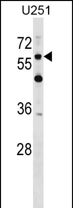
CSPG5 Antibody (C-term)
Affinity Purified Rabbit Polyclonal Antibody (Pab)
- SPECIFICATION
- CITATIONS
- PROTOCOLS
- BACKGROUND

Application
| WB, E |
|---|---|
| Primary Accession | O95196 |
| Other Accession | NP_006565.2 |
| Reactivity | Human |
| Host | Rabbit |
| Clonality | Polyclonal |
| Isotype | Rabbit IgG |
| Calculated MW | 60016 Da |
| Antigen Region | 530-559 aa |
| Gene ID | 10675 |
|---|---|
| Other Names | Chondroitin sulfate proteoglycan 5, Acidic leucine-rich EGF-like domain-containing brain protein, Neuroglycan C, CSPG5, CALEB, NGC |
| Target/Specificity | This CSPG5 antibody is generated from rabbits immunized with a KLH conjugated synthetic peptide between 530-559 amino acids from the C-terminal region of human CSPG5. |
| Dilution | WB~~1:1000 E~~Use at an assay dependent concentration. |
| Format | Purified polyclonal antibody supplied in PBS with 0.09% (W/V) sodium azide. This antibody is purified through a protein A column, followed by peptide affinity purification. |
| Storage | Maintain refrigerated at 2-8°C for up to 2 weeks. For long term storage store at -20°C in small aliquots to prevent freeze-thaw cycles. |
| Precautions | CSPG5 Antibody (C-term) is for research use only and not for use in diagnostic or therapeutic procedures. |
| Name | CSPG5 |
|---|---|
| Synonyms | CALEB, NGC |
| Function | May function as a growth and differentiation factor involved in neuritogenesis. May induce ERBB3 activation. |
| Cellular Location | Cell membrane {ECO:0000250|UniProtKB:Q9ERQ6}; Single-pass type I membrane protein {ECO:0000250|UniProtKB:Q9ERQ6} Synaptic cell membrane {ECO:0000250|UniProtKB:Q71M36}; Single-pass type I membrane protein {ECO:0000250|UniProtKB:Q71M36}. Endoplasmic reticulum membrane {ECO:0000250|UniProtKB:Q71M36}; Single-pass type I membrane protein {ECO:0000250|UniProtKB:Q71M36}. Golgi apparatus membrane {ECO:0000250|UniProtKB:Q71M36}; Single-pass type I membrane protein {ECO:0000250|UniProtKB:Q71M36}. Cell surface {ECO:0000250|UniProtKB:Q71M36}. Secreted. Note=In neurons, localizes to synaptic junctions. Also detected in the endoplasmic reticulum and the Golgi Partially enriched in lipid rafts. {ECO:0000250|UniProtKB:Q71M36, ECO:0000250|UniProtKB:Q9ERQ6} |
| Tissue Location | Detected in cerebrospinal fluid (at protein level) (PubMed:25326458). Detected in urine (at protein level) (PubMed:37453717). Expressed in brain (at protein level) (PubMed:9950058). |

Thousands of laboratories across the world have published research that depended on the performance of antibodies from Abcepta to advance their research. Check out links to articles that cite our products in major peer-reviewed journals, organized by research category.
info@abcepta.com, and receive a free "I Love Antibodies" mug.

Provided below are standard protocols that you may find useful for product applications.
Background
CSPG5 may function as a growth and differentiation factor involved in neuritogenesis. May induce ERBB3 activation.
References
So, H.C., et al. Am. J. Med. Genet. B Neuropsychiatr. Genet. 153B (1), 103-113 (2010) :
Ichihara-Tanaka, K., et al. J. Biol. Chem. 281(41):30857-30864(2006)
Aono, S., et al. J. Neurosci. Res. 83(1):110-118(2006)
Kinugasa, Y., et al. Biochem. Biophys. Res. Commun. 321(4):1045-1049(2004)
Hassel, B., et al. J. Biol. Chem. 278(41):40136-40143(2003)
If you have used an Abcepta product and would like to share how it has performed, please click on the "Submit Review" button and provide the requested information. Our staff will examine and post your review and contact you if needed.
If you have any additional inquiries please email technical services at tech@abcepta.com.
Ordering Information
Other Products
Shipping Information
