


 Foundational characteristics of cancer include proliferation, angiogenesis, migration, evasion of apoptosis, and cellular immortality. Find key markers for these cellular processes and antibodies to detect them.
 The SUMOplot™ Analysis Program predicts and scores sumoylation sites in your protein. SUMOylation is a post-translational modification involved in various cellular processes, such as nuclear-cytosolic transport, transcriptional regulation, apoptosis, protein stability, response to stress, and progression through the cell cycle.
 The Autophagy Receptor Motif Plotter predicts and scores autophagy receptor binding sites in your protein. Identifying proteins connected to this pathway is critical to understanding the role of autophagy in physiological as well as pathological processes such as development, differentiation, neurodegenerative diseases, stress, infection, and cancer.
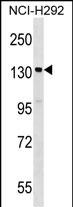
SIPA1 Antibody (C-term)
Affinity Purified Rabbit Polyclonal Antibody (Pab)
- SPECIFICATION
- CITATIONS
- PROTOCOLS
- BACKGROUND

Application
| WB, E |
|---|---|
| Primary Accession | Q96FS4 |
| Other Accession | NP_006738.3, NP_694985.29 |
| Reactivity | Human |
| Host | Rabbit |
| Clonality | Polyclonal |
| Isotype | Rabbit IgG |
| Calculated MW | 112149 Da |
| Antigen Region | 942-970 aa |
| Gene ID | 6494 |
|---|---|
| Other Names | Signal-induced proliferation-associated protein 1, Sipa-1, GTPase-activating protein Spa-1, p130 SPA-1, SIPA1, SPA1 |
| Target/Specificity | This SIPA1 antibody is generated from rabbits immunized with a KLH conjugated synthetic peptide between 942-970 amino acids from the C-terminal region of human SIPA1. |
| Dilution | WB~~1:1000 E~~Use at an assay dependent concentration. |
| Format | Purified polyclonal antibody supplied in PBS with 0.09% (W/V) sodium azide. This antibody is purified through a protein A column, followed by peptide affinity purification. |
| Storage | Maintain refrigerated at 2-8°C for up to 2 weeks. For long term storage store at -20°C in small aliquots to prevent freeze-thaw cycles. |
| Precautions | SIPA1 Antibody (C-term) is for research use only and not for use in diagnostic or therapeutic procedures. |
| Name | SIPA1 |
|---|---|
| Synonyms | SPA1 |
| Function | GTPase activator for the nuclear Ras-related regulatory proteins Rap1 and Rap2 in vitro, converting them to the putatively inactive GDP-bound state (PubMed:9346962). Affects cell cycle progression (By similarity). |
| Cellular Location | Nucleus. Cytoplasm, perinuclear region. Endomembrane system; Peripheral membrane protein. Note=Mostly localized in the perinuclear membraneous region |
| Tissue Location | Expressed in fetal as well as in adult tissues. Expressed abundantly in the lymphoid tissues such as thymus, spleen and peripheral blood lymphocytes and also shows a significant expression in the spinal cord |

Thousands of laboratories across the world have published research that depended on the performance of antibodies from Abcepta to advance their research. Check out links to articles that cite our products in major peer-reviewed journals, organized by research category.
info@abcepta.com, and receive a free "I Love Antibodies" mug.

Provided below are standard protocols that you may find useful for product applications.
Background
The product of this gene is a mitogen induced GTPase activating protein (GAP). It exhibits a specific GAP activity for Ras-related regulatory proteins Rap1 and Rap2, but not for Ran or other small GTPases. This protein may also hamper mitogen-induced cell cycle progression when abnormally or prematurely expressed. It is localized to the perinuclear region. Two alternatively spliced variants encoding the same isoform have been characterized to date.
References
Bailey, S.D., et al. Diabetes Care 33(10):2250-2253(2010)
Brooks, R., et al. Gynecol. Oncol. 116(3):539-543(2010)
Hosgood, H.D. III, et al. Occup Environ Med 66(12):848-853(2009)
Talmud, P.J., et al. Am. J. Hum. Genet. 85(5):628-642(2009)
Hsieh, S.M., et al. Breast Cancer Res. 11 (5), R75 (2009) :
If you have used an Abcepta product and would like to share how it has performed, please click on the "Submit Review" button and provide the requested information. Our staff will examine and post your review and contact you if needed.
If you have any additional inquiries please email technical services at tech@abcepta.com.
Ordering Information
Other Products
Shipping Information
