


 Foundational characteristics of cancer include proliferation, angiogenesis, migration, evasion of apoptosis, and cellular immortality. Find key markers for these cellular processes and antibodies to detect them.
 The SUMOplot™ Analysis Program predicts and scores sumoylation sites in your protein. SUMOylation is a post-translational modification involved in various cellular processes, such as nuclear-cytosolic transport, transcriptional regulation, apoptosis, protein stability, response to stress, and progression through the cell cycle.
 The Autophagy Receptor Motif Plotter predicts and scores autophagy receptor binding sites in your protein. Identifying proteins connected to this pathway is critical to understanding the role of autophagy in physiological as well as pathological processes such as development, differentiation, neurodegenerative diseases, stress, infection, and cancer.
ESX1 Antibody (N-term)
Affinity Purified Rabbit Polyclonal Antibody (Pab)
- SPECIFICATION
- CITATIONS
- PROTOCOLS
- BACKGROUND

Application
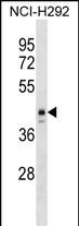
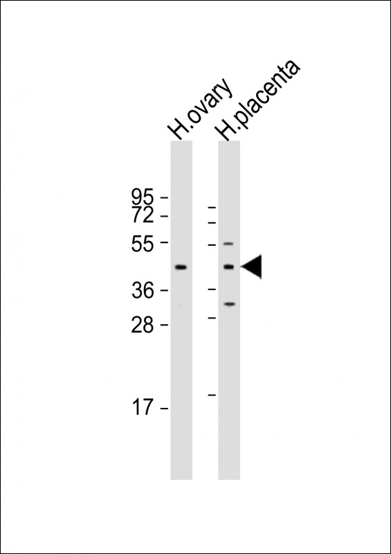
| WB, E |
|---|---|
| Primary Accession | Q8N693 |
| Other Accession | NP_703149.1 |
| Reactivity | Human |
| Host | Rabbit |
| Clonality | Polyclonal |
| Isotype | Rabbit IgG |
| Calculated MW | 44297 Da |
| Antigen Region | 28-55 aa |
| Gene ID | 80712 |
|---|---|
| Other Names | Homeobox protein ESX1, Extraembryonic, spermatogenesis, homeobox 1, Homeobox protein ESX1-N, Homeobox protein ESX1-C, ESX1, ESX1L, ESX1R |
| Target/Specificity | This ESX1 antibody is generated from rabbits immunized with a KLH conjugated synthetic peptide between 28-55 amino acids from the N-terminal region of human ESX1. |
| Dilution | WB~~1:1000-1:2000 E~~Use at an assay dependent concentration. |
| Format | Purified polyclonal antibody supplied in PBS with 0.09% (W/V) sodium azide. This antibody is purified through a protein A column, followed by peptide affinity purification. |
| Storage | Maintain refrigerated at 2-8°C for up to 2 weeks. For long term storage store at -20°C in small aliquots to prevent freeze-thaw cycles. |
| Precautions | ESX1 Antibody (N-term) is for research use only and not for use in diagnostic or therapeutic procedures. |
| Name | ESX1 |
|---|---|
| Synonyms | ESX1L, ESX1R |
| Function | May coordinately regulate cell cycle progression and transcription during spermatogenesis. Inhibits degradation of polyubiquitinated cyclin A and cyclin B1 and thereby arrests the cell cycle at early M phase. ESXR1-N acts as a transcriptional repressor. Binds to the sequence 5'-TAATGTTATTA-3' which is present within the first intron of the KRAS gene and inhibits its expression. ESXR1-C has the ability to inhibit cyclin turnover. |
| Cellular Location | Cytoplasm. Nucleus {ECO:0000255|PROSITE-ProRule:PRU00108, ECO:0000269|PubMed:15235584} Note=ESXR1-N localizes specifically to the nucleus while ESXR1-C localizes specifically to the cytoplasm |
| Tissue Location | Expressed in placenta and testis. Expressed in testicular germ cell tumors. |

Thousands of laboratories across the world have published research that depended on the performance of antibodies from Abcepta to advance their research. Check out links to articles that cite our products in major peer-reviewed journals, organized by research category.
info@abcepta.com, and receive a free "I Love Antibodies" mug.

Provided below are standard protocols that you may find useful for product applications.
Background
This gene encodes a dual-function 65 kDa protein that undergoes proteolytic cleavage to produce a 45 kDa N-terminal fragment with a paired-like homeodomain and a 20 kDa C-terminal fragment with a proline-rich domain. The C-terminal fragment localizes to the cytoplasm while the N-terminal fragment localizes exclusively to the nucleus. In contrast to human, the mouse homolog has a novel PN/PF motif in the C-terminus and is paternally imprinted in placental tissue. This gene likely plays a role in placental development and spermatogenesis.
References
Bonaparte, E., et al. Hum. Reprod. 25(6):1398-1403(2010)
Wang, X., et al. Hum. Mol. Genet. 16(17):2053-2060(2007)
Loschiavo, M., et al. Mamm. Genome 18(1):75-85(2007)
Murthi, P., et al. Mol. Hum. Reprod. 12(5):335-340(2006)
Yanagihara, M., et al. Oncogene 24(38):5878-5887(2005)
If you have used an Abcepta product and would like to share how it has performed, please click on the "Submit Review" button and provide the requested information. Our staff will examine and post your review and contact you if needed.
If you have any additional inquiries please email technical services at tech@abcepta.com.
Ordering Information
Other Products
Shipping Information
