


 Foundational characteristics of cancer include proliferation, angiogenesis, migration, evasion of apoptosis, and cellular immortality. Find key markers for these cellular processes and antibodies to detect them.
 The SUMOplot™ Analysis Program predicts and scores sumoylation sites in your protein. SUMOylation is a post-translational modification involved in various cellular processes, such as nuclear-cytosolic transport, transcriptional regulation, apoptosis, protein stability, response to stress, and progression through the cell cycle.
 The Autophagy Receptor Motif Plotter predicts and scores autophagy receptor binding sites in your protein. Identifying proteins connected to this pathway is critical to understanding the role of autophagy in physiological as well as pathological processes such as development, differentiation, neurodegenerative diseases, stress, infection, and cancer.
NPLOC4 Antibody (C-term)
Affinity Purified Rabbit Polyclonal Antibody (Pab)
- SPECIFICATION
- CITATIONS
- PROTOCOLS
- BACKGROUND

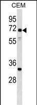
Application
| WB, E |
|---|---|
| Primary Accession | Q8TAT6 |
| Other Accession | Q9ES54, P60670, NP_060391.2 |
| Reactivity | Human |
| Predicted | Mouse, Rat |
| Host | Rabbit |
| Clonality | Polyclonal |
| Isotype | Rabbit IgG |
| Calculated MW | 68120 Da |
| Antigen Region | 456-484 aa |
| Gene ID | 55666 |
|---|---|
| Other Names | Nuclear protein localization protein 4 homolog, Protein NPL4, NPLOC4, KIAA1499, NPL4 |
| Target/Specificity | This NPLOC4 antibody is generated from rabbits immunized with a KLH conjugated synthetic peptide between 456-484 amino acids from the C-terminal region of human NPLOC4. |
| Dilution | WB~~1:1000 E~~Use at an assay dependent concentration. |
| Format | Purified polyclonal antibody supplied in PBS with 0.09% (W/V) sodium azide. This antibody is purified through a protein A column, followed by peptide affinity purification. |
| Storage | Maintain refrigerated at 2-8°C for up to 2 weeks. For long term storage store at -20°C in small aliquots to prevent freeze-thaw cycles. |
| Precautions | NPLOC4 Antibody (C-term) is for research use only and not for use in diagnostic or therapeutic procedures. |
| Name | NPLOC4 |
|---|---|
| Synonyms | KIAA1499, NPL4 |
| Function | The ternary complex containing UFD1, VCP and NPLOC4 binds ubiquitinated proteins and is necessary for the export of misfolded proteins from the ER to the cytoplasm, where they are degraded by the proteasome. The NPLOC4-UFD1-VCP complex regulates spindle disassembly at the end of mitosis and is necessary for the formation of a closed nuclear envelope (By similarity). Acts as a negative regulator of type I interferon production via the complex formed with VCP and UFD1, which binds to RIGI and recruits RNF125 to promote ubiquitination and degradation of RIGI (PubMed:26471729). |
| Cellular Location | Cytoplasm, cytosol {ECO:0000250|UniProtKB:Q9ES54}. Endoplasmic reticulum {ECO:0000250|UniProtKB:Q9ES54}. Nucleus {ECO:0000250|UniProtKB:Q9ES54} Note=Associated with the endoplasmic reticulum and nuclear {ECO:0000250|UniProtKB:Q9ES54} |
| Tissue Location | Expressed at highest levels in brain, heart, skeletal muscle, kidney and fetal liver. |

Thousands of laboratories across the world have published research that depended on the performance of antibodies from Abcepta to advance their research. Check out links to articles that cite our products in major peer-reviewed journals, organized by research category.
info@abcepta.com, and receive a free "I Love Antibodies" mug.

Provided below are standard protocols that you may find useful for product applications.
Background
The ternary complex containing UFD1L, VCP and NPLOC4 binds ubiquitinated proteins and is necessary for the export of misfolded proteins from the ER to the cytoplasm, where they are degraded by the proteasome. The NPLOC4-UFD1L-VCP complex regulates spindle disassembly at the end of mitosis and is necessary for the formation of a closed nuclear envelope (By similarity).
References
Liu, F., et al. PLoS Genet. 6, E1000934 (2010) :
Lass, A., et al. Exp. Cell Res. 314(14):2715-2723(2008)
McConnell, E., et al. Biochem. Biophys. Res. Commun. 355(4):1087-1090(2007)
Gevaert, K., et al. Nat. Biotechnol. 21(5):566-569(2003)
Botta, A., et al. Gene 275(1):39-46(2001)
If you have used an Abcepta product and would like to share how it has performed, please click on the "Submit Review" button and provide the requested information. Our staff will examine and post your review and contact you if needed.
If you have any additional inquiries please email technical services at tech@abcepta.com.
Ordering Information
Other Products
Shipping Information
