


 Foundational characteristics of cancer include proliferation, angiogenesis, migration, evasion of apoptosis, and cellular immortality. Find key markers for these cellular processes and antibodies to detect them.
 The SUMOplot™ Analysis Program predicts and scores sumoylation sites in your protein. SUMOylation is a post-translational modification involved in various cellular processes, such as nuclear-cytosolic transport, transcriptional regulation, apoptosis, protein stability, response to stress, and progression through the cell cycle.
 The Autophagy Receptor Motif Plotter predicts and scores autophagy receptor binding sites in your protein. Identifying proteins connected to this pathway is critical to understanding the role of autophagy in physiological as well as pathological processes such as development, differentiation, neurodegenerative diseases, stress, infection, and cancer.
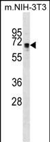
NDOR1 Antibody (Center)
Affinity Purified Rabbit Polyclonal Antibody (Pab)
- SPECIFICATION
- CITATIONS
- PROTOCOLS
- BACKGROUND

Application
| WB, E |
|---|---|
| Primary Accession | Q9UHB4 |
| Other Accession | Q1JPJ0, NP_055249.1, NP_001137498.1 |
| Reactivity | Human, Mouse |
| Predicted | Bovine |
| Host | Rabbit |
| Clonality | Polyclonal |
| Isotype | Rabbit IgG |
| Calculated MW | 66763 Da |
| Antigen Region | 207-234 aa |
| Gene ID | 27158 |
|---|---|
| Other Names | NADPH-dependent diflavin oxidoreductase 1 {ECO:0000255|HAMAP-Rule:MF_03178}, 16-- {ECO:0000255|HAMAP-Rule:MF_03178}, NADPH-dependent FMN and FAD-containing oxidoreductase {ECO:0000255|HAMAP-Rule:MF_03178}, Novel reductase 1, NDOR1 {ECO:0000255|HAMAP-Rule:MF_03178}, NR1 |
| Target/Specificity | This NDOR1 antibody is generated from rabbits immunized with a KLH conjugated synthetic peptide between 207-234 amino acids from the Central region of human NDOR1. |
| Dilution | WB~~1:1000 E~~Use at an assay dependent concentration. |
| Format | Purified polyclonal antibody supplied in PBS with 0.09% (W/V) sodium azide. This antibody is purified through a protein A column, followed by peptide affinity purification. |
| Storage | Maintain refrigerated at 2-8°C for up to 2 weeks. For long term storage store at -20°C in small aliquots to prevent freeze-thaw cycles. |
| Precautions | NDOR1 Antibody (Center) is for research use only and not for use in diagnostic or therapeutic procedures. |
| Name | NDOR1 {ECO:0000255|HAMAP-Rule:MF_03178} |
|---|---|
| Function | NADPH-dependent reductase which is a central component of the cytosolic iron-sulfur (Fe-S) protein assembly (CIA) machinery (PubMed:10625700, PubMed:15900210, PubMed:20802492, PubMed:23596212, PubMed:28648056). Transfers electrons from NADPH via its FAD and FMN prosthetic groups to the [2Fe-2S] cluster of CIAPIN1, another key component of the CIA machinery (PubMed:20802492, PubMed:23596212, PubMed:28648056). In turn, this reduced cluster provides electrons for assembly of cytosolic iron-sulfur cluster proteins (PubMed:20802492, PubMed:23596212). It can also reduce the [2Fe-2S] cluster of CISD1 and activate this protein implicated in Fe/S cluster repair (PubMed:28648056). In vitro can fully activate methionine synthase/MTR in the presence of soluble cytochrome b5/CYB5A (PubMed:12871938). |
| Cellular Location | Cytoplasm, perinuclear region {ECO:0000255|HAMAP- Rule:MF_03178, ECO:0000269|PubMed:10625700, ECO:0000269|PubMed:12871939}. Note=Concentrated in perinuclear structure. {ECO:0000255|HAMAP-Rule:MF_03178, ECO:0000269|PubMed:12871939} |
| Tissue Location | Low expression in brain, heart, kidney, pancreas, prostate and skeletal muscle. Highest levels in the placenta. Expressed in cancer cell lines including promyelocytic leukemia, HeLaS3, chronic myelagenous leukemia, lymphoblastic leukemia, Burkitt's lymphoma, colorectal adenocarcinoma, lung carcinoma, and melanoma G-361 |

Thousands of laboratories across the world have published research that depended on the performance of antibodies from Abcepta to advance their research. Check out links to articles that cite our products in major peer-reviewed journals, organized by research category.
info@abcepta.com, and receive a free "I Love Antibodies" mug.

Provided below are standard protocols that you may find useful for product applications.
Background
This gene encodes an NADPH-dependent diflavin reductase that contains both flavin mononucleotide (FMN) and flavin adenine dinucleotide (FAD) binding domains. The encoded protein is an enzyme that catalyzes the transfers electrons from NADPH through FAD and FMN cofactors to potential redox partners. Alternative splicing results in multiple transcript variants. [provided by RefSeq].
References
Kwasnicka-Crawford, D.A., et al. Biochem. Biophys. Res. Commun. 336(2):565-571(2005)
Finn, R.D., et al. Pharmacogenet. Genomics 15(6):381-386(2005)
Kwasnicka, D.A., et al. J. Biol. Chem. 278(40):39051-39058(2003)
Olteanu, H., et al. J. Biol. Chem. 278(40):38310-38314(2003)
Finn, R.D., et al. Eur. J. Biochem. 270(6):1164-1175(2003)
If you have used an Abcepta product and would like to share how it has performed, please click on the "Submit Review" button and provide the requested information. Our staff will examine and post your review and contact you if needed.
If you have any additional inquiries please email technical services at tech@abcepta.com.
Ordering Information
Other Products
Shipping Information
