


 Foundational characteristics of cancer include proliferation, angiogenesis, migration, evasion of apoptosis, and cellular immortality. Find key markers for these cellular processes and antibodies to detect them.
 The SUMOplot™ Analysis Program predicts and scores sumoylation sites in your protein. SUMOylation is a post-translational modification involved in various cellular processes, such as nuclear-cytosolic transport, transcriptional regulation, apoptosis, protein stability, response to stress, and progression through the cell cycle.
 The Autophagy Receptor Motif Plotter predicts and scores autophagy receptor binding sites in your protein. Identifying proteins connected to this pathway is critical to understanding the role of autophagy in physiological as well as pathological processes such as development, differentiation, neurodegenerative diseases, stress, infection, and cancer.
NDOR1 Polyclonal Antibody
Purified Rabbit Polyclonal Antibody (Pab)
- SPECIFICATION
- CITATIONS
- PROTOCOLS
- BACKGROUND

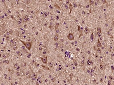
Application
| IHC-P, IHC-F, IF, ICC |
|---|---|
| Primary Accession | Q9UHB4 |
| Reactivity | Rat, Pig, Dog, Bovine |
| Host | Rabbit |
| Clonality | Polyclonal |
| Calculated MW | 67 KDa |
| Physical State | Liquid |
| Immunogen | KLH conjugated synthetic peptide derived from human NDOR1 |
| Epitope Specificity | 51-150/597 |
| Isotype | IgG |
| Purity | affinity purified by Protein A |
| Buffer | 0.01M TBS (pH7.4) with 1% BSA, 0.02% Proclin300 and 50% Glycerol. |
| SUBCELLULAR LOCATION | Cytoplasm > perinuclear region. Concentrated in perinuclear structure. |
| SIMILARITY | In the C-terminal section; belongs to the flavoprotein pyridine nucleotide cytochrome reductase family. Contains 1 FAD-binding FR-type domain. Contains 1 flavodoxin-like domain. |
| Important Note | This product as supplied is intended for research use only, not for use in human, therapeutic or diagnostic applications. |
| Background Descriptions | This gene encodes an NADPH-dependent diflavin reductase that contains both flavin mononucleotide (FMN) and flavin adenine dinucleotide (FAD) binding domains. The encoded protein catalyzes the transfer of electrons from NADPH through FAD and FMN cofactors to potential redox partners. Alternative splicing results in multiple transcript variants. [provided by RefSeq, Mar 2012] |
| Gene ID | 27158 |
|---|---|
| Other Names | NADPH-dependent diflavin oxidoreductase 1 {ECO:0000255|HAMAP-Rule:MF_03178}, 1.18.1.- {ECO:0000255|HAMAP-Rule:MF_03178}, NADPH-dependent FMN and FAD-containing oxidoreductase {ECO:0000255|HAMAP-Rule:MF_03178}, Novel reductase 1, NDOR1 {ECO:0000255|HAMAP-Rule:MF_03178}, NR1 |
| Target/Specificity | Low expression in brain, heart, kidney, pancreas, prostate and skeletal muscle. Highest levels in the placenta. Expressed in cancer cell lines including promyelocytic leukemia, HeLa S3, chronic myelagenous leukemia, lymphoblastic leukemia, Burkitt's lymphoma, colorectal adenocarcinoma, lung carcinoma, and melanoma G361. |
| Dilution | IHC-P=1:100-500,IHC-F=1:100-500,ICC=1:100-500,IF=1:100-500 |
| Storage | Store at -20 ℃ for one year. Avoid repeated freeze/thaw cycles. When reconstituted in sterile pH 7.4 0.01M PBS or diluent of antibody the antibody is stable for at least two weeks at 2-4 ℃. |
| Name | NDOR1 {ECO:0000255|HAMAP-Rule:MF_03178} |
|---|---|
| Function | NADPH-dependent reductase which is a central component of the cytosolic iron-sulfur (Fe-S) protein assembly (CIA) machinery (PubMed:10625700, PubMed:15900210, PubMed:20802492, PubMed:23596212, PubMed:28648056). Transfers electrons from NADPH via its FAD and FMN prosthetic groups to the [2Fe-2S] cluster of CIAPIN1, another key component of the CIA machinery (PubMed:20802492, PubMed:23596212, PubMed:28648056). In turn, this reduced cluster provides electrons for assembly of cytosolic iron-sulfur cluster proteins (PubMed:20802492, PubMed:23596212). It can also reduce the [2Fe-2S] cluster of CISD1 and activate this protein implicated in Fe/S cluster repair (PubMed:28648056). In vitro can fully activate methionine synthase/MTR in the presence of soluble cytochrome b5/CYB5A (PubMed:12871938). |
| Cellular Location | Cytoplasm, perinuclear region {ECO:0000255|HAMAP- Rule:MF_03178, ECO:0000269|PubMed:10625700, ECO:0000269|PubMed:12871939}. Note=Concentrated in perinuclear structure. {ECO:0000255|HAMAP-Rule:MF_03178, ECO:0000269|PubMed:12871939} |
| Tissue Location | Low expression in brain, heart, kidney, pancreas, prostate and skeletal muscle. Highest levels in the placenta. Expressed in cancer cell lines including promyelocytic leukemia, HeLaS3, chronic myelagenous leukemia, lymphoblastic leukemia, Burkitt's lymphoma, colorectal adenocarcinoma, lung carcinoma, and melanoma G-361 |
Research Areas
Citations (0)

Thousands of laboratories across the world have published research that depended on the performance of antibodies from Abcepta to advance their research. Check out links to articles that cite our products in major peer-reviewed journals, organized by research category.
Submit your citation using an Abcepta antibody to
info@abcepta.com, and receive a free "I Love Antibodies" mug.
info@abcepta.com, and receive a free "I Love Antibodies" mug.

Application Protocols
Provided below are standard protocols that you may find useful for product applications.
Abcepta welcomes feedback from its customers.
If you have used an Abcepta product and would like to share how it has performed, please click on the "Submit Review" button and provide the requested information. Our staff will examine and post your review and contact you if needed.
If you have any additional inquiries please email technical services at tech@abcepta.com.
$ 385.00
Cat# AP57377
Ordering Information
United States
AlbaniaAustraliaAustriaBelgiumBosnia & HerzegovinaBrazilBulgariaCanadaCentral AmericaChinaCroatiaCyprusCzech RepublicDenmarkEstoniaFinlandFranceGermanyGreeceHong KongHungaryIcelandIndiaIndonesiaIrelandIsraelItalyJapanLatviaLithuaniaLuxembourgMacedoniaMalaysiaMaltaMexicoNetherlandsNew ZealandNorwayPakistanPolandPortugalRomaniaSerbiaSingaporeSlovakiaSloveniaSouth AfricaSouth KoreaSpainSwedenSwitzerlandTaiwanTurkeyUnited KingdomUnited StatesVietnamWorldwideOthers
USA Headquarters
(888) 735-7227 / (858) 622-0099 or (858) 875-1900
Other Products
Shipping Information
Domestic orders (in stock items)
Shipped out the same day. Orders placed after 1 PM (PST) will ship out the next business day.
International orders
Contact your local distributors
