


 Foundational characteristics of cancer include proliferation, angiogenesis, migration, evasion of apoptosis, and cellular immortality. Find key markers for these cellular processes and antibodies to detect them.
 The SUMOplot™ Analysis Program predicts and scores sumoylation sites in your protein. SUMOylation is a post-translational modification involved in various cellular processes, such as nuclear-cytosolic transport, transcriptional regulation, apoptosis, protein stability, response to stress, and progression through the cell cycle.
 The Autophagy Receptor Motif Plotter predicts and scores autophagy receptor binding sites in your protein. Identifying proteins connected to this pathway is critical to understanding the role of autophagy in physiological as well as pathological processes such as development, differentiation, neurodegenerative diseases, stress, infection, and cancer.
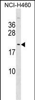
TTDN1 Antibody (C-term)
Affinity Purified Rabbit Polyclonal Antibody (Pab)
- SPECIFICATION
- CITATIONS
- PROTOCOLS
- BACKGROUND

Application
| WB, E |
|---|---|
| Primary Accession | Q8TAP9 |
| Other Accession | Q9D011, NP_619646.1 |
| Reactivity | Human |
| Predicted | Mouse |
| Host | Rabbit |
| Clonality | Polyclonal |
| Isotype | Rabbit IgG |
| Calculated MW | 19147 Da |
| Antigen Region | 151-177 aa |
| Gene ID | 136647 |
|---|---|
| Other Names | M-phase-specific PLK1-interacting protein, TTD non-photosensitive 1 protein, MPLKIP, C7orf11, TTDN1 |
| Target/Specificity | This TTDN1 antibody is generated from rabbits immunized with a KLH conjugated synthetic peptide between 151-177 amino acids from the C-terminal region of human TTDN1. |
| Dilution | WB~~1:1000 E~~Use at an assay dependent concentration. |
| Format | Purified polyclonal antibody supplied in PBS with 0.09% (W/V) sodium azide. This antibody is purified through a protein A column, followed by peptide affinity purification. |
| Storage | Maintain refrigerated at 2-8°C for up to 2 weeks. For long term storage store at -20°C in small aliquots to prevent freeze-thaw cycles. |
| Precautions | TTDN1 Antibody (C-term) is for research use only and not for use in diagnostic or therapeutic procedures. |
| Name | MPLKIP |
|---|---|
| Synonyms | C7orf11, TTDN1 |
| Function | May play a role in maintenance of cell cycle integrity by regulating mitosis or cytokinesis. |
| Cellular Location | Nucleus. Cytoplasm. Cytoplasm, cytoskeleton, microtubule organizing center, centrosome. Note=The subcellular location is regulated during cell cycle. During interphase located in the nucleus. During mitosis located at the centrosome and dispersed in the cytoplasm. During telophase located in the midbody. Colocalizes with PLK1 at the centrosome in M phase |
| Tissue Location | Expressed at highest levels in liver and kidney; intermediate expression in skeletal muscle, pancreas, heart and placenta; low expression in brain and lung. Expressed in epidermis and hair follicles. |

Thousands of laboratories across the world have published research that depended on the performance of antibodies from Abcepta to advance their research. Check out links to articles that cite our products in major peer-reviewed journals, organized by research category.
info@abcepta.com, and receive a free "I Love Antibodies" mug.

Provided below are standard protocols that you may find useful for product applications.
Background
The protein encoded by this gene localizes to the centrosome during mitosis and to the midbody during cytokinesis. The protein is phosphorylated by cyclin-dependent kinase 1 during mitosis and subsequently interacts with polo-like kinase 1. The protein is thought to function in regulating mitosis and cytokinesis. Mutations in this gene result in nonphotosensitive trichothiodystrophy.
References
Zhang, Y., et al. Cell. Mol. Life Sci. 64(5):632-640(2007)
Lamesch, P., et al. Genomics 89(3):307-315(2007)
Botta, E., et al. Hum. Mutat. 28(1):92-96(2007)
Olsen, J.V., et al. Cell 127(3):635-648(2006)
Nakabayashi, K., et al. Am. J. Hum. Genet. 76(3):510-516(2005)
If you have used an Abcepta product and would like to share how it has performed, please click on the "Submit Review" button and provide the requested information. Our staff will examine and post your review and contact you if needed.
If you have any additional inquiries please email technical services at tech@abcepta.com.
Ordering Information
Other Products
Shipping Information
