


 Foundational characteristics of cancer include proliferation, angiogenesis, migration, evasion of apoptosis, and cellular immortality. Find key markers for these cellular processes and antibodies to detect them.
 The SUMOplot™ Analysis Program predicts and scores sumoylation sites in your protein. SUMOylation is a post-translational modification involved in various cellular processes, such as nuclear-cytosolic transport, transcriptional regulation, apoptosis, protein stability, response to stress, and progression through the cell cycle.
 The Autophagy Receptor Motif Plotter predicts and scores autophagy receptor binding sites in your protein. Identifying proteins connected to this pathway is critical to understanding the role of autophagy in physiological as well as pathological processes such as development, differentiation, neurodegenerative diseases, stress, infection, and cancer.
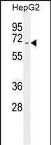
UAP1 Antibody (C-term)
Affinity Purified Rabbit Polyclonal Antibody (Pab)
- SPECIFICATION
- CITATIONS
- PROTOCOLS
- BACKGROUND

Application
| WB, E |
|---|---|
| Primary Accession | Q16222 |
| Other Accession | Q91YN5, NP_003106.3 |
| Reactivity | Human |
| Predicted | Mouse |
| Host | Rabbit |
| Clonality | Polyclonal |
| Isotype | Rabbit IgG |
| Calculated MW | 58769 Da |
| Antigen Region | 390-418 aa |
| Gene ID | 6675 |
|---|---|
| Other Names | UDP-N-acetylhexosamine pyrophosphorylase, Antigen X, AGX, Sperm-associated antigen 2, UDP-N-acetylgalactosamine pyrophosphorylase, AGX-1, UDP-N-acetylglucosamine pyrophosphorylase, AGX-2, UAP1, SPAG2 |
| Target/Specificity | This UAP1 antibody is generated from rabbits immunized with a KLH conjugated synthetic peptide between 390-418 amino acids from the C-terminal region of human UAP1. |
| Dilution | WB~~1:1000 E~~Use at an assay dependent concentration. |
| Format | Purified polyclonal antibody supplied in PBS with 0.09% (W/V) sodium azide. This antibody is purified through a protein A column, followed by peptide affinity purification. |
| Storage | Maintain refrigerated at 2-8°C for up to 2 weeks. For long term storage store at -20°C in small aliquots to prevent freeze-thaw cycles. |
| Precautions | UAP1 Antibody (C-term) is for research use only and not for use in diagnostic or therapeutic procedures. |
| Name | UAP1 {ECO:0000303|PubMed:9603950, ECO:0000312|HGNC:HGNC:12457} |
|---|---|
| Function | Catalyzes the last step in biosynthesis of uridine diphosphate-N-acetylglucosamine (UDP-GlcNAc) by converting UTP and glucosamine 1-phosphate (GlcNAc-1-P) to the sugar nucleotide (PubMed:9603950, PubMed:9765219). Also converts UTP and galactosamine 1-phosphate (GalNAc-1-P) into uridine diphosphate-N-acetylgalactosamine (UDP-GalNAc) (PubMed:9765219). In addition to its role in metabolism, acts as a regulator of innate immunity in response to virus infection by mediating pyrophosphorylation of IRF3: catalyzes pyrophosphorylation of IRF3 phosphorylated at 'Ser-386' by TBK1, promoting IRF3 dimerization and activation, leading to type I interferon responses (PubMed:36603579). |
| Cellular Location | Cytoplasm, cytosol. Note=In spermatozoa, localized to the principal piece of the tail, the neck region of the head and to a lesser extent, the midpiece of the tail. |
| Tissue Location | Widely expressed (PubMed:8025165). Expressed at low level in placenta, muscle and liver (PubMed:8025165) [Isoform AGX2]: Isoform AGX2 is more abundant than isoform AGX1 in somatic tissue. |

Thousands of laboratories across the world have published research that depended on the performance of antibodies from Abcepta to advance their research. Check out links to articles that cite our products in major peer-reviewed journals, organized by research category.
info@abcepta.com, and receive a free "I Love Antibodies" mug.

Provided below are standard protocols that you may find useful for product applications.
Background
UAP1 converts UDP and GlcNAc-1-P into UDP-GlcNAc, and UDP and GalNAc-1-P into UDP-GalNAc. Isoform AGX1 has 2 to 3 times higher activity towards GalNAc-1-P, while isoform AGX2 has 8 times more activity towards GlcNAc-1-P.
References
Ehret, G.B., et al. Eur. J. Hum. Genet. 17(12):1650-1657(2009)
Wang, A.G., et al. Biochem. Biophys. Res. Commun. 345(3):1022-1032(2006)
Peneff, C., et al. EMBO J. 20(22):6191-6202(2001)
Wang-Gillam, A., et al. J. Biol. Chem. 273(42):27055-27057(1998)
Diekman, A.B., et al. Mol. Reprod. Dev. 50(3):284-293(1998)
If you have used an Abcepta product and would like to share how it has performed, please click on the "Submit Review" button and provide the requested information. Our staff will examine and post your review and contact you if needed.
If you have any additional inquiries please email technical services at tech@abcepta.com.
Ordering Information
Other Products
Shipping Information
