


 Foundational characteristics of cancer include proliferation, angiogenesis, migration, evasion of apoptosis, and cellular immortality. Find key markers for these cellular processes and antibodies to detect them.
 The SUMOplot™ Analysis Program predicts and scores sumoylation sites in your protein. SUMOylation is a post-translational modification involved in various cellular processes, such as nuclear-cytosolic transport, transcriptional regulation, apoptosis, protein stability, response to stress, and progression through the cell cycle.
 The Autophagy Receptor Motif Plotter predicts and scores autophagy receptor binding sites in your protein. Identifying proteins connected to this pathway is critical to understanding the role of autophagy in physiological as well as pathological processes such as development, differentiation, neurodegenerative diseases, stress, infection, and cancer.
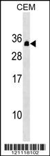
PDCD10 Antibody (Center)
Affinity Purified Rabbit Polyclonal Antibody (Pab)
- SPECIFICATION
- CITATIONS
- PROTOCOLS
- BACKGROUND

Application
| WB, E |
|---|---|
| Primary Accession | Q9BUL8 |
| Other Accession | Q8AVR4, Q6NX65, Q8VE70, Q5ZIV5, Q6PHH3, NP_009148.2 |
| Reactivity | Human |
| Predicted | Zebrafish, Chicken, Mouse, Rat, Xenopus |
| Host | Rabbit |
| Clonality | Polyclonal |
| Isotype | Rabbit IgG |
| Calculated MW | 24702 Da |
| Antigen Region | 96-124 aa |
| Gene ID | 11235 |
|---|---|
| Other Names | Programmed cell death protein 10, Cerebral cavernous malformations 3 protein, TF-1 cell apoptosis-related protein 15, PDCD10, CCM3, TFAR15 |
| Target/Specificity | This PDCD10 antibody is generated from rabbits immunized with a KLH conjugated synthetic peptide between 96-124 amino acids from the Central region of human PDCD10. |
| Dilution | WB~~1:1000 E~~Use at an assay dependent concentration. |
| Format | Purified polyclonal antibody supplied in PBS with 0.09% (W/V) sodium azide. This antibody is purified through a protein A column, followed by peptide affinity purification. |
| Storage | Maintain refrigerated at 2-8°C for up to 2 weeks. For long term storage store at -20°C in small aliquots to prevent freeze-thaw cycles. |
| Precautions | PDCD10 Antibody (Center) is for research use only and not for use in diagnostic or therapeutic procedures. |
| Name | PDCD10 (HGNC:8761) |
|---|---|
| Function | Promotes cell proliferation. Modulates apoptotic pathways. Increases mitogen-activated protein kinase activity and STK26 activity (PubMed:27807006). Important for cell migration, and for normal structure and assembly of the Golgi complex (PubMed:27807006). Part of the striatin-interacting phosphatase and kinase (STRIPAK) complexes. STRIPAK complexes have critical roles in protein (de)phosphorylation and are regulators of multiple signaling pathways including Hippo, MAPK, nuclear receptor and cytoskeleton remodeling. Different types of STRIPAK complexes are involved in a variety of biological processes such as cell growth, differentiation, apoptosis, metabolism and immune regulation (PubMed:18782753). Important for KDR/VEGFR2 signaling. Increases the stability of KDR/VEGFR2 and prevents its breakdown. Required for normal cardiovascular development. Required for normal angiogenesis, vasculogenesis and hematopoiesis during embryonic development (By similarity). |
| Cellular Location | Cytoplasm. Golgi apparatus membrane {ECO:0000250|UniProtKB:Q6NX65}; Peripheral membrane protein {ECO:0000250|UniProtKB:Q6NX65}; Cytoplasmic side {ECO:0000250|UniProtKB:Q6NX65}. Cell membrane {ECO:0000250|UniProtKB:Q6NX65}; Peripheral membrane protein {ECO:0000250|UniProtKB:Q6NX65}; Cytoplasmic side {ECO:0000250|UniProtKB:Q6NX65} |
| Tissue Location | Ubiquitous.. |

Thousands of laboratories across the world have published research that depended on the performance of antibodies from Abcepta to advance their research. Check out links to articles that cite our products in major peer-reviewed journals, organized by research category.
info@abcepta.com, and receive a free "I Love Antibodies" mug.

Provided below are standard protocols that you may find useful for product applications.
Background
This gene encodes an evolutionarily conserved protein associated with cell apoptosis. The protein interacts with the serine/threonine protein kinase MST4 to modulate the extracellular signal-regulated kinase (ERK) pathway. It also interacts with and is phosphoryated by serine/threonine kinase 25, and is thought to function in a signaling pathway essential for vascular developent. Mutations in this gene are one cause of cerebral cavernous malformations, which are vascular malformations that cause seizures and cerebral hemorrhages. Multiple alternatively spliced variants, encoding the same protein, have been identified. [provided by RefSeq].
References
Lauenborg, B., et al. APMIS 118(10):719-728(2010)
Shimada, M., et al. Hum. Genet. 128(4):433-441(2010)
Ding, J., et al. Biochem. Biophys. Res. Commun. 399(4):587-592(2010)
Zheng, X., et al. J. Clin. Invest. 120(8):2795-2804(2010)
Dibble, C.F., et al. PLoS ONE 5 (7), E11740 (2010) :
If you have used an Abcepta product and would like to share how it has performed, please click on the "Submit Review" button and provide the requested information. Our staff will examine and post your review and contact you if needed.
If you have any additional inquiries please email technical services at tech@abcepta.com.
Ordering Information
Shipping Information
