


 Foundational characteristics of cancer include proliferation, angiogenesis, migration, evasion of apoptosis, and cellular immortality. Find key markers for these cellular processes and antibodies to detect them.
 The SUMOplot™ Analysis Program predicts and scores sumoylation sites in your protein. SUMOylation is a post-translational modification involved in various cellular processes, such as nuclear-cytosolic transport, transcriptional regulation, apoptosis, protein stability, response to stress, and progression through the cell cycle.
 The Autophagy Receptor Motif Plotter predicts and scores autophagy receptor binding sites in your protein. Identifying proteins connected to this pathway is critical to understanding the role of autophagy in physiological as well as pathological processes such as development, differentiation, neurodegenerative diseases, stress, infection, and cancer.
PDCD10 Polyclonal Antibody
Purified Rabbit Polyclonal Antibody (Pab)
- SPECIFICATION
- CITATIONS
- PROTOCOLS
- BACKGROUND

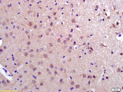
Application
| IHC-P, IHC-F, IF, E |
|---|---|
| Primary Accession | Q9BUL8 |
| Reactivity | Rat, Pig |
| Host | Rabbit |
| Clonality | Polyclonal |
| Calculated MW | 22 KDa |
| Physical State | Liquid |
| Immunogen | KLH conjugated synthetic peptide derived from human PDCD10 |
| Epitope Specificity | 145-212/212 |
| Isotype | IgG |
| Purity | affinity purified by Protein A |
| Buffer | 0.01M TBS (pH7.4) with 1% BSA, 0.02% Proclin300 and 50% Glycerol. |
| SUBCELLULAR LOCATION | Cytoplasm. Golgi apparatus membrane; Peripheral membrane protein; Cytoplasmic side. Cell membrane; Peripheral membrane protein; Cytoplasmic side. Note=Partially co-localizes with endogenous PXN at the leading edges of migrating cells. |
| SIMILARITY | Belongs to the PDCD10 family. |
| SUBUNIT | Homodimer. Interacts (via C-terminus) with CCM2 and PXN. Interacts (via N-terminus) with MST4, STK24 and STK25. Interacts with GOLGA2. Identified in a complex with CCM1 and CCM2. Interacts with KDR/VEGFR2. Interaction with KDR/VEGFR2 is enhanced by stimulation with VEGFA. |
| DISEASE | Defects in PDCD10 are the cause of cerebral cavernous malformations type 3 (CCM3) [MIM:603285]. Cerebral cavernous malformations (CCMs) are congenital vascular anomalies of the central nervous system that can result in hemorrhagic stroke, seizures, recurrent headaches, and focal neurologic deficits. CCMs have an incidence of 0.1%-0.5% in the general population and usually present clinically during the 3rd to 5th decade of life. The lesions are characterized by grossly enlarged blood vessels consisting of a single layer of endothelium and without any intervening neural tissue, ranging in diameter from a few millimeters to several centimeters. |
| Important Note | This product as supplied is intended for research use only, not for use in human, therapeutic or diagnostic applications. |
| Background Descriptions | This gene encodes an evolutionarily conserved protein associated with cell apoptosis. The protein interacts with the serine/threonine protein kinase MST4 to modulate the extracellular signal-regulated kinase (ERK) pathway. It also interacts with and is phosphoryated by serine/threonine kinase 25, and is thought to function in a signaling pathway essential for vascular developent. Mutations in this gene are one cause of cerebral cavernous malformations, which are vascular malformations that cause seizures and cerebral hemorrhages. Multiple alternatively spliced variants, encoding the same protein, have been identified. [provided by RefSeq, Jul 2008]. |
| Gene ID | 11235 |
|---|---|
| Other Names | Programmed cell death protein 10, Cerebral cavernous malformations 3 protein, TF-1 cell apoptosis-related protein 15, PDCD10, CCM3, TFAR15 |
| Target/Specificity | Ubiquitous. |
| Dilution | IHC-P=1:100-500,IHC-F=1:100-500,IF=1:100-500,ELISA=1:5000-10000 |
| Storage | Store at -20 ℃ for one year. Avoid repeated freeze/thaw cycles. When reconstituted in sterile pH 7.4 0.01M PBS or diluent of antibody the antibody is stable for at least two weeks at 2-4 ℃. |
| Name | PDCD10 (HGNC:8761) |
|---|---|
| Function | Promotes cell proliferation. Modulates apoptotic pathways. Increases mitogen-activated protein kinase activity and STK26 activity (PubMed:27807006). Important for cell migration, and for normal structure and assembly of the Golgi complex (PubMed:27807006). Part of the striatin-interacting phosphatase and kinase (STRIPAK) complexes. STRIPAK complexes have critical roles in protein (de)phosphorylation and are regulators of multiple signaling pathways including Hippo, MAPK, nuclear receptor and cytoskeleton remodeling. Different types of STRIPAK complexes are involved in a variety of biological processes such as cell growth, differentiation, apoptosis, metabolism and immune regulation (PubMed:18782753). Important for KDR/VEGFR2 signaling. Increases the stability of KDR/VEGFR2 and prevents its breakdown. Required for normal cardiovascular development. Required for normal angiogenesis, vasculogenesis and hematopoiesis during embryonic development (By similarity). |
| Cellular Location | Cytoplasm. Golgi apparatus membrane {ECO:0000250|UniProtKB:Q6NX65}; Peripheral membrane protein {ECO:0000250|UniProtKB:Q6NX65}; Cytoplasmic side {ECO:0000250|UniProtKB:Q6NX65}. Cell membrane {ECO:0000250|UniProtKB:Q6NX65}; Peripheral membrane protein {ECO:0000250|UniProtKB:Q6NX65}; Cytoplasmic side {ECO:0000250|UniProtKB:Q6NX65} |
| Tissue Location | Ubiquitous.. |
Research Areas
Citations (0)

Thousands of laboratories across the world have published research that depended on the performance of antibodies from Abcepta to advance their research. Check out links to articles that cite our products in major peer-reviewed journals, organized by research category.
Submit your citation using an Abcepta antibody to
info@abcepta.com, and receive a free "I Love Antibodies" mug.
info@abcepta.com, and receive a free "I Love Antibodies" mug.

Application Protocols
Provided below are standard protocols that you may find useful for product applications.
Abcepta welcomes feedback from its customers.
If you have used an Abcepta product and would like to share how it has performed, please click on the "Submit Review" button and provide the requested information. Our staff will examine and post your review and contact you if needed.
If you have any additional inquiries please email technical services at tech@abcepta.com.
$ 385.00
Cat# AP58526
Ordering Information
United States
AlbaniaAustraliaAustriaBelgiumBosnia & HerzegovinaBrazilBulgariaCanadaCentral AmericaChinaCroatiaCyprusCzech RepublicDenmarkEstoniaFinlandFranceGermanyGreeceHong KongHungaryIcelandIndiaIndonesiaIrelandIsraelItalyJapanLatviaLithuaniaLuxembourgMacedoniaMalaysiaMaltaMexicoNetherlandsNew ZealandNorwayPakistanPolandPortugalRomaniaSerbiaSingaporeSlovakiaSloveniaSouth AfricaSouth KoreaSpainSwedenSwitzerlandTaiwanTurkeyUnited KingdomUnited StatesVietnamWorldwideOthers
USA Headquarters
(888) 735-7227 / (858) 622-0099 or (858) 875-1900
Shipping Information
Domestic orders (in stock items)
Shipped out the same day. Orders placed after 1 PM (PST) will ship out the next business day.
International orders
Contact your local distributors
