


 Foundational characteristics of cancer include proliferation, angiogenesis, migration, evasion of apoptosis, and cellular immortality. Find key markers for these cellular processes and antibodies to detect them.
 The SUMOplot™ Analysis Program predicts and scores sumoylation sites in your protein. SUMOylation is a post-translational modification involved in various cellular processes, such as nuclear-cytosolic transport, transcriptional regulation, apoptosis, protein stability, response to stress, and progression through the cell cycle.
 The Autophagy Receptor Motif Plotter predicts and scores autophagy receptor binding sites in your protein. Identifying proteins connected to this pathway is critical to understanding the role of autophagy in physiological as well as pathological processes such as development, differentiation, neurodegenerative diseases, stress, infection, and cancer.
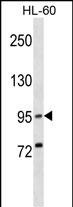
XPR1 Antibody (N-term)
Affinity Purified Rabbit Polyclonal Antibody (Pab)
- SPECIFICATION
- CITATIONS
- PROTOCOLS
- BACKGROUND

Application
| WB, E |
|---|---|
| Primary Accession | Q9UBH6 |
| Other Accession | Q9Z0U0, Q9QZ70, NP_001129141.1 |
| Reactivity | Human |
| Predicted | Hamster, Mouse |
| Host | Rabbit |
| Clonality | Polyclonal |
| Isotype | Rabbit IgG |
| Calculated MW | 81535 Da |
| Antigen Region | 96-122 aa |
| Gene ID | 9213 |
|---|---|
| Other Names | Xenotropic and polytropic retrovirus receptor 1, Protein SYG1 homolog, Xenotropic and polytropic murine leukemia virus receptor X3, X-receptor, XPR1, SYG1, XR |
| Target/Specificity | This XPR1 antibody is generated from rabbits immunized with a KLH conjugated synthetic peptide between 96-122 amino acids from the N-terminal region of human XPR1. |
| Dilution | WB~~1:1000 E~~Use at an assay dependent concentration. |
| Format | Purified polyclonal antibody supplied in PBS with 0.09% (W/V) sodium azide. This antibody is purified through a protein A column, followed by peptide affinity purification. |
| Storage | Maintain refrigerated at 2-8°C for up to 2 weeks. For long term storage store at -20°C in small aliquots to prevent freeze-thaw cycles. |
| Precautions | XPR1 Antibody (N-term) is for research use only and not for use in diagnostic or therapeutic procedures. |
| Name | XPR1 {ECO:0000303|PubMed:31043717} |
|---|---|
| Function | Inorganic ion transporter that mediates phosphate ion export across plasma membrane (PubMed:23791524, PubMed:25938945, PubMed:27080106, PubMed:31043717, PubMed:39169184, PubMed:39325866, PubMed:39747008, PubMed:39814721). Plays a major role in phosphate homeostasis, preventing intracellular phosphate accumulation and possible calcium phosphate precipitation, ultimately preserving calcium signaling (PubMed:27080106). Binds inositol hexakisphosphate (Ins6P) and similar inositol polyphosphates, such as 5-diphospho-inositol pentakisphosphate (5-InsP7), which are important intracellular signaling molecules involved in regulation of phosphate flux (PubMed:27080106, PubMed:39169184, PubMed:39325866). |
| Cellular Location | Cell membrane; Multi-pass membrane protein |
| Tissue Location | Widely expressed. Detected in spleen, lymph node, thymus, leukocytes, bone marrow, heart, kidney, pancreas and skeletal muscle. |

Thousands of laboratories across the world have published research that depended on the performance of antibodies from Abcepta to advance their research. Check out links to articles that cite our products in major peer-reviewed journals, organized by research category.
info@abcepta.com, and receive a free "I Love Antibodies" mug.

Provided below are standard protocols that you may find useful for product applications.
Background
XPR1 may function in G-protein coupled signal transduction (By similarity). Potential receptor for xenotropic and polytropic murine leukemia retroviruses.
References
Bhosle, S., et al. J. Virol. 84(13):6288-6296(2010)
Rose, J.E., et al. Mol. Med. 16 (7-8), 247-253 (2010) :
Battini, J.L., et al. Proc. Natl. Acad. Sci. U.S.A. 96(4):1385-1390(1999)
Tailor, C.S., et al. Proc. Natl. Acad. Sci. U.S.A. 96(3):927-932(1999)
Yang, Y.L., et al. Nat. Genet. 21(2):216-219(1999)
If you have used an Abcepta product and would like to share how it has performed, please click on the "Submit Review" button and provide the requested information. Our staff will examine and post your review and contact you if needed.
If you have any additional inquiries please email technical services at tech@abcepta.com.
Ordering Information
Other Products
Shipping Information
