


 Foundational characteristics of cancer include proliferation, angiogenesis, migration, evasion of apoptosis, and cellular immortality. Find key markers for these cellular processes and antibodies to detect them.
 The SUMOplot™ Analysis Program predicts and scores sumoylation sites in your protein. SUMOylation is a post-translational modification involved in various cellular processes, such as nuclear-cytosolic transport, transcriptional regulation, apoptosis, protein stability, response to stress, and progression through the cell cycle.
 The Autophagy Receptor Motif Plotter predicts and scores autophagy receptor binding sites in your protein. Identifying proteins connected to this pathway is critical to understanding the role of autophagy in physiological as well as pathological processes such as development, differentiation, neurodegenerative diseases, stress, infection, and cancer.
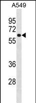
FCRL1 Antibody (C-term)
Affinity Purified Rabbit Polyclonal Antibody (Pab)
- SPECIFICATION
- CITATIONS
- PROTOCOLS
- BACKGROUND

Application
| WB, E |
|---|---|
| Primary Accession | Q96LA6 |
| Other Accession | NP_001152869.1 |
| Reactivity | Human |
| Host | Rabbit |
| Clonality | Polyclonal |
| Isotype | Rabbit IgG |
| Calculated MW | 46936 Da |
| Antigen Region | 366-392 aa |
| Gene ID | 115350 |
|---|---|
| Other Names | Fc receptor-like protein 1, FcR-like protein 1, FcRL1, Fc receptor homolog 1, FcRH1, IFGP family protein 1, hIFGP1, Immune receptor translocation-associated protein 5, CD307a, FCRL1, FCRH1, IFGP1, IRTA5 |
| Target/Specificity | This FCRL1 antibody is generated from rabbits immunized with a KLH conjugated synthetic peptide between 366-392 amino acids from the C-terminal region of human FCRL1. |
| Dilution | WB~~1:1000 E~~Use at an assay dependent concentration. |
| Format | Purified polyclonal antibody supplied in PBS with 0.09% (W/V) sodium azide. This antibody is purified through a protein A column, followed by peptide affinity purification. |
| Storage | Maintain refrigerated at 2-8°C for up to 2 weeks. For long term storage store at -20°C in small aliquots to prevent freeze-thaw cycles. |
| Precautions | FCRL1 Antibody (C-term) is for research use only and not for use in diagnostic or therapeutic procedures. |
| Name | FCRL1 |
|---|---|
| Synonyms | FCRH1, IFGP1, IRTA5 |
| Function | Type I transmembrane surface glycoprotein preferentially expressed by B-cells that regulates BCR-mediated signaling responses (PubMed:15479727). Recruits ABL1 as the intracellular effector molecule to enhance B-cell activation (By similarity). Also plays a negative role by suppressing ERK activation under homeostatic and BCR-stimulated conditions in a GRB2-dependent manner (By similarity). |
| Cellular Location | Cell membrane; Single-pass type I membrane protein |
| Tissue Location | Primarily expressed in secondary lymphoid tissues by mature subsets of B-cells. Detected in spleen, lymph node, heart, skeletal muscle, kidney, liver and placenta. Specifically expressed by mature B lineage cells with higher expression in naive versus memory B- cells (at protein level). |

Thousands of laboratories across the world have published research that depended on the performance of antibodies from Abcepta to advance their research. Check out links to articles that cite our products in major peer-reviewed journals, organized by research category.
info@abcepta.com, and receive a free "I Love Antibodies" mug.

Provided below are standard protocols that you may find useful for product applications.
Background
This gene encodes a member of the immunoglobulin receptor superfamily and is one of several Fc receptor-like glycoproteins clustered on the long arm of chromosome 1. The encoded protein contains three extracellular C2-like immunoglobulin domains, a transmembrane domain and a cytoplasmic domain with two immunoreceptor-tyrosine activation motifs. This protein may play a role in the regulation of cancer cell growth. Alternative splicing results in multiple transcript variants.
References
Rose, J.E., et al. Mol. Med. 16 (7-8), 247-253 (2010) :
Davila, S., et al. Genes Immun. 11(3):232-238(2010)
Kazemi, T., et al. Cancer Immunol. Immunother. 58(6):989-996(2009)
Kazemi, T., et al. Int. J. Cancer 123(9):2113-2119(2008)
Du, X., et al. Blood 111(1):338-343(2008)
If you have used an Abcepta product and would like to share how it has performed, please click on the "Submit Review" button and provide the requested information. Our staff will examine and post your review and contact you if needed.
If you have any additional inquiries please email technical services at tech@abcepta.com.
Ordering Information
Other Products
Shipping Information
